|
|
Case Reports Indian Pediatrics 2001; 38: 553-556 |
|||||
|
Giant Congenital Melanocytic Nevus with Cutis Verticis Gyrata |
|||||
|
Giant congenital melanocytic nevi (GCMN), bathing trunk nevi or garment nevi represent a rare group of pigmentary naevi covering large areas of the body surface. We are reporting a case of GCMN in a neonate because of the unusually large extent of the lesion, its presentation over the scalp as cutis verticis gyrata (CVG) and its association with Nevus of Ota in the mother.
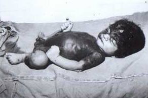
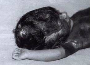
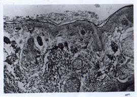
A six-day-old neonate, born of non-consanguineous marriage by normal vaginal delivery at term to a primigravida with uneventful antenatal history presented with an extensive pigmented patch over the body since birth. Physical examination revealed an exten-sive bluish-black patch covering 80% of the skin surface including most of the head and neck, trunk, buttocks and proximal aspects of the upper and lower limbs. Multiple pigmented satellite lesions were also present over the extremities. Tufts of coarse and lusterless hair were scattered over the lesions. There was a tendency for verrucous plaque formation over the frontal and temporal regions of the scalp and upper arms (Fig. 1). The lesion on the scalp was hypertrophied and thrown into longi-tudinal folds indicative of CVG (Fig. 2). Fundus examination, X-ray spine, USG cranium and abdomen were normal. Skin biopsy taken from the scalp revealed presence of polygonal nevoid cells in the upper dermis containing melanin. Scattered nests of sebaceous glands were also present. No junctional activity or evidence of maligant tranformation was seen (Fig. 3). The mother had a violaceous patch over the right peri-orbital region and sclera suggestive of Nevus of Ota.
Congenital melanocytic nevi are pig-mented cutaneous lesions formed by a combination of epidermally and dermally derived naevus cells occuring in about one per cent of the newborns(1). They are arbitrarily classified according to their size as small (<1.5 cm), medium (1.5 - 19.9 cm) and large or giant nevi (>20 cm)(2). GCMN occur in approximately 1 of 20,000 live births(3). Clini-cally, they are characterized by large blue-black patches presenting at birth commonly over the back and thigh area, with smaller darker patches interspersed as satellite lesions. The giant nevi involving the head and neck may be associated with meningeal melano-cytosis, sometimes complicated by hydro-cephalus and malignant melanoma. Lesions over the back in midline are associated with spina bifida(4). Several studies in the past have suggested a malignant potential of these lesions with the incidence ranging from 2-41%(5). Also, about 40% of the malignant melanomas in children arise within these CGMN(6). GCMN are usually known to cover 20-30% of the skin surface(7). However, our patient presented with an unusually, extensive GCMN covering 80% of the body surface area including almost the entire head and neck region. The lesion over the scalp was morphologically typical of CVG. The term ‘CVG’ describes the hypertrophy and folding of the skin of the scalp to present a gyrate or cerebriform appearance. Etiologically, this morphology may either have a genetic predisposition or occurs secondary to pachydermoperiostosis, various endocrine disorders like acromegaly and myxoedema, neurofibroms or melanocytic nevi(8). In a series of 85 cases of diseases of scalp in the form of CVG, 24 cases (21%) were secondary to neoplasms, all of which were nevi (ectodermal or mesodermal in origin). Though most of the reported cases have been nevi of melanocytic type, but neurofibromas and fibromas can assume this form(9). To the best of our knowledge, no association between GCMN and Nevus of Ota has been reported(5). However in our case, the mother had Nevus of Ota and the baby presented with GCMN. GCMN are cosmetically very distressing to the parents. Also, the significant risk of malignant change warrants prophylactic removal. However their large size poses a great surgical challenge. The object of the treatment is to remove as many as possible of the melanocytic nevus cells by ablative approaches, thus improve both the cosmetic appearance and reduce the risk of malignant melanoma. This may be achieved by total excision of the nevus followed by resurfacing with autografts. Dermabrasion has also been successfully employed with gratifying cosmetic results, but leaving behind of nevus cells in the deep dermis has been a serious objection to the procedure(4,6). Contributors: NBM and AM interpreted the findings and finalized the draft. SK interpreted the dermato-logical findings and drafted the paper. SB helped in drafting the paper. Funding: None. Competing interests: None stated.
Fig. 1. Giant congenital hairy melanocytic nevus with CVG.
Fig. 2. Scalp lesion thrown into longitudinal folds suggestive of CVG.
|
![]()