|
|
Case Reports Indian Pediatrics 2001; 38: 543-545 |
|||||
|
Non Hodgkins Lymphoma Presenting as Cardiac Tumor |
|||||
|
Lymphoma is known to be the third most common cancer in children and non-Hodgkin’s lymphoma (NHL), one of its two broad categories represents the malignant clonal proliferations of primarily T or B-lymphocytes. NHL is about 1.5 times more common than Hodgkin’s lymphoma in young children and it affects boys three times as frequently as it does girls. The clinical features of NHL depends upon the site of the tumor and the extent of the disease e.g., in the head neck region it present as painless enlargement of lymph nodes; in the abdomen as mass or intestinal obstruction; in the CNS as increased intracranial pressure. In the chest it usually arises in the anterior mediastinum leading to compression of the airways and superior vena cava. Very rarely it may affect the heart and present as a cardiac tumor. We are reporting such a case where NHL presented as a cardiac tumor.
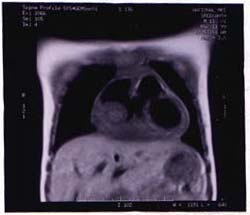
An 11-year-old boy was brought with chest pain and intermittent low-grade fever of two weeks duration. It was a dull aching pain which increased with inspiration and was relieved by sitting up. He had no other symptoms like dyspnea, arthritis, rashes, etc. At six years of age this boy had acute rheumatic fever with carditis and pericardial effusion, according to the medical records. He recovered from it fully and was on regular penicillin prophylaxis since then. Physical examination at the time of admission showed a well-grown child with a pulse rate of 100 per minute with normal rhythm, volume and character; respiratory rate of 22/min and BP of 120/72 mm of Hg. he had no pallor, cyanosis, edema or significant lymphadenopathy at any of the lymph node areas. CVS examination showed prominent suprasternal pulsations with an elevated and pulsatile JVP 6 cm above the sternal angle. The heart sounds were normal and there was no cardiomegaly or murmurs at the time of admission. There was tender hepatomegaly of 4 cm with no splenomegaly or other palpable masses in the abdomen. With these findings, the initial provisional diagnostic possibilities considered were rheumatic recurrence with carditis and congestive cardiac failure, and tuberculous pericarditis. Investigations revealed an ESR of 110 mm/h with a normal TLC, DLC, Hb and peripheral smear. Mantoux test and ASO titre were negative. X-ray chest showed cardiomegaly with prominence of right atrium with a cardio-thoracic ratio of 60% and ECG showed right bundle branch block with T inversion in leads L II and AVF. One week after admission, dyspnea increased and a diastolic murmur appeared at the tricuspid area. A repeat chest X-ray was taken which showed that CT ratio had increased from 60 to 70%. An echocardiogram was done which showed a large mass in the right atrium attached to its lateral wall. MRI confirmed the same mass with extension through the pericardium, suggestive of a malignant tumor (Fig. 1). After thoracic surgery consultation, considering a primary cardiac tumor, he was taken up for exploratory surgery the next week. Per operatively a hard nodular mass infiltrating the whole of the right atrial wall (except the appendages) extending through the pericardium to the right hilum was seen. In view of extensive nature of the lesion, tumor resection was deferred and multiple biopsies taken. Post operatively, the child developed resistant arrhythmia and hypotension and died in the same evening. Later the biopsy report came as Non Hodgkin’s lymphoma of lymphoblastic type.
Fig. 1. MRI scan showing mass in the right atrium.
NHL of childhood is usually a diffuse extranodal high-grade tumor. According to the National Cancer Institute of USA. it is divided into three subtypes namely, the small non-cleaved cell, lymphoblastic and large cell types. The most frequent primary sites are the abdomen (31.4%), the head and neck region (29%) and the mediastinum (26%). The non-cervical lymph nodes involvement is usually 6.5% while skin, thyroid, epidural space and bone accounts for the remainder 7%(1). The anterior mediastinum is usually affected by the lymphoblastic type and it commonly presents as pleural effusion, respiratory distress or superior venacaval syndrome. The heart, as in this case, is described as one of the rare sites of primary lymphoma along with the other sites like the orbit, breast, salivary gland, and adrenal gland(2). After an Internet search, we were able to find out only a few case reports of primary cardiac lymphoma of which only two were pediatric cases(3,4). Mediastinal and heart involvement is usually evaluated by chest X-ray, echocardiogram, CT scan, MRI and rarely by mediastinography and thoracotomy. Tissue diagnosis is established by FNAC or open biopsy. Once the diagnosis is established, accurate staging is essential before start of therapy. It is divided into 4 stages according to Murphy(5) and the treatment depends on the stage of the disease. The mainstay of treatment is multiagent chemotherapy. The commonly used therapies are the cyclophosphamide based COMP regimen and the intensive multi-agent LSA2-L2 regimen(6). With modern therapy, most cases of NHL have good prognosis, especially if diagnosed early. The two-year event-free survival is approximately 90% with limited stage diseases and 70% for those with Stage III or IV disease. This case shows how a relatively common and curable disease like NHL could present in an uncommon manner leading to problems in early diagnosis and institution of specific therapy. Contributors: BR collected data and prepared the manuscript. He will act as the guarantor for the paper. KN and CMA helped in drafting and revision. Funding: None. Competing interests: None stated.
|
![]()