Jatinder S.
Goraya
P.N. Gupta*
Ravi K. Gupta*
Raj Bahadur*
V.R. Parmar
From the Departments of Pediatrics and
Orthopedics*, Government Medical College and Hospital,
Chandigarh 160 047, India.
Reprint requests: Jatinder S. Goraya, Senior
Lecturer, Department of Pediatrics, Government Medical College
and Hospital, Chandigarh 160 047, India.
Manuscript Received: March 22, 1999;
Initial review completed: May 12, 1999;
Revision Accepted: August 19, 1999
Epilepsy is a common neurological disorder of
childhood frequently requiring prolonged use of anticonvulsants.
Most of the commonly used anticonvulsants particularly phenytoin,
pheno-barbitone, and carbamazepine have the propensity to
interfere with vitamin D meta-bolism. Though biochemical changes
are frequent, clinically overt rickets or osteomalacia is
rare(1-6). Since the signs and symptoms of osteomalacia are
non-specific, the diagnosis is frequently delayed resulting in
considerable morbidity(1,2). We report three cases with
anticonvulsant induced osteomalacia.
Case Reports
Case 1:
A 15-year-old adolescent girl
presented with pain above the left knee and limp of 1½-month
duration. She had received various analgesic drugs without any
relief. She was a known epileptic, having recurrent seizures
despite being on carbamazepine (26 mg/kg/day) and phenobarbitone
(3 mg/kg/day) for the last 2½ and 1 year, respectively. Because
of poor seizure control, she was spending most of her time
indoors. About one year prior to presenta-tion, she was diagnosed
to have pulmonary tuberculosis and had received anti-tuberculous
drugs (2 HRZE + 4HR) for 6 months. She had been complaining of
non-specific body aches and pains with generalized weakness for
the last 6 months. No complaints indicative of malabsorption,
liver, or renal disease were present.
Examination revealed tenderness over left
distal femoral shaft and an antalgic gait. Rest of the examination
was unremarkable. X-ray left knee revealed a fracture line
in the distal femoral diaphysis (Fig. 1). The patient was
suspected to have anticonvulsants induced osteomalacia. A skeletal
survey was perofrmed which showed Looser’s zones affecting ribs,
and pubic ramus (Fig. 2) and generalized decrease in bone
density. In addition, radiological evidence of rickets was present
in the form of metaphyseal fraying and widening of epiphyseal
plates. Serum biochemistry revealed serum calcium of 7.8 mg/dL
(normal 8.2-11.5 mg/dL), phosporus 3.5 mg/dL (normal 2.7-4.8 mg/dL)
and a raised alkaline phosphatase of 1491 U/L (normal 66-279.0
U/L). She was treated with single oral dose of 6,00,000 units of
vitamin D and plaster immobilization for 6 weeks. Patient showed
significant subjective improvement in her symptoms. By 8 weeks of
treatment she was fully ambulatory and had minimal limp and normal
serum calcium.
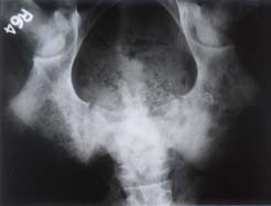 |
Fig. 1.
Radiograph showing fracture line in
the distal femoral diaphysis. |
Case 2:
A 17-year-old adolescent female
presented with backache and limp of 6 months duration following a
seizure related fall. X-rays done outside revealed
insufficiency fractures of both femoral necks. She was treated
with traction, analgesics and calcium supplements without any
relief; rather her symptoms worsened and she was unable to walk.
Examination revealed tenderness and limitation of movements at
both hips. Serum biochemistry revealed low serum calcium of 8.1
mg/dL, phosphorus 3.5 mg/dL and raised alkaline phosphatase of
1020 U/L. X-ray pelvis revealed insufficiency fracture in
both femoral necks. She was treated with an oral dose of 6,00,000
units of vitamin D and immobilization for 6 weeks with skin
traction. She was a known epileptic receiving phenobarbitone (2
mg/kg/day) and carbamazepine (14 mg/kg/day) for the last 5 and 3
years, respectively. Serum calcium normalized by 6 weeks of
treatment and alkaline phos-phatase started decreasing (773 U/L).
The insufficiency fracture in both femoral necks healed and she
became ambulatory.
| Fig. 2.
Radiograph showing Looser’s zone in the pubic ramus |
|
.
Case 3:
A 7-month-old boy was noticed to
have widening of wrists, costochondral beading, and frontal
bossing.
|
.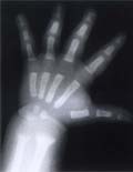 |
X-ray of the left wrist revealed rachitic changes
(Fig. 3). He was receiving phenobarbitone (4 mg/kg/day)
since 3 months of age for seizures, which he had developed in
association with pyogenic menin-gitis. He was on formula feeds
since birth and was weaned at the age of 6 months. There was no
history to suggest chronic diarrhea or lack of exposure to
sunlight. He weighed 7.5 kg and had a head circumference of 43 cm.
Systemic examination was unremarkable. Serum bioc-hemistry
revealed calcium 10.0 mg/dL, phos-phorous 3.8 mg/dL, and a raised
alkaline phosphatase of 1293 U/L. Patient was treated with a
single dose of 600,000 units of vitamin D. Repeat X-ray
left wrist done after 3 weeks showed evidence of healing rickets. |
|
Fig. 3.
Radiograph of the wrist showing rachitic changes |
Discussion
Anticonvulsants induced osteomalacia is
uncommonly recognized complication of long term anti-epileptic
medication(1,2). The apparent association between long-term
anticonvulsant therapy and biochemical and radiological changes of
rickets was first described by Kruse in 1968 from Germany(7) and
later by Dent et al. in 1970 from England(8).
The frequency of problem is related to the
population of patients studied and the method of detection
employed(1). Majority of the studies have been done in
institutionalized, non-ambulatory patients receiving high dose
multiple anticonvulsants for several years with sub-optimal
dietary intake of vitamin D as well as limited exposure to
sunlight(1-4,6-9). Biochemical evidence of osteomalacia, i.e.,
alteration in serum calcium, phosphorus and alkaline phosphatase
has been seen in 20-70% of these pateints. Radiological changes of
osteomalacia have been detected in upto 50% of cases(1-6).
Clinically overt osteomalacia is, however rare compared to
biochemical and radiological changes(1,2,4). Since the clinical
features of osteomalacia, viz., musculoskeletal pains,
weakness are nonspecific, the diagnosis is easily over-looked as
happened in our patients(Cases I and II). Rarely, occurrence of
fractures, both seizure and non-seizure related, lead to the
correct diagnosis as happened in two of our patients (Cases I and
II).
Anticonvulsants exert their deleterious effects
on mineral and bone metabolism by inducing hepatic microsomal
mixed function oxidase enzyme systems, resulting in increased
catabolism and excretion of vitamin D and its biologically active
products(1,2). In addition, drugs especially phenytoin may
directly inhibit the intestinal calcium absorption(1).
Several factors enhance the risk of
anticonvulsants induced bone disease(1,2). Patient related factors
include decreased dietary vitamin D, limited exposure to sunlight,
reduced physical activity(1,3,10-12). The latter two factors were
present in one of our patient (Case I). In addition all our
patients were in the age which corresponded to the period of rapid
growth, i.e., adolescence (Cases I and II) and infancy
(Case III). Rapid growth might have contributed to the depletion
of already vulnerable vitamin D stores of the patients(3).
Though all commonly used anticonvulsants have
the propensity to cause osteomalacia, phenytoin and phenobarbitone
have the greatest potential(1-3). Most of the patients, who
develop anticonvulsant induced osteomalacia, usually are on these
drugs for several years though osteomalacia has also been
described to set in within 3-6 months of therapy(13,14). Patients
who receive more than one drug are at greater risk than those who
are on mono-therapy(3,4,9). Apart from being on multiple durgs, i.e.,
carbamazepine and phenobarbitone, one of our patients (Case I)
also had received rifampicin, which is another drug, which induces
hepatic enzymes and interferes with vitamin D metabolism. It may
be argued that Case III had nutritional rickets which are
otherwise common in infancy. But in the presence of adequate
nutritional status and lack of any other risk factors,
phenobarbitone was thought to have contributed to a large extent
in the development of rickets in this infant.
Diagnosis of anticonvulsant induced
osteomalacia involves exclusion of other causes of osteomalacia
particularly malabsorption, renal and hepatic disease(2) of which
there was no evidence in our patients. The diagnosis should be
considered in epileptic patients receiving anticonvulsant drugs,
who develop bone pains, muscle weakness, fractures after minimal
trauma or have worsening of seizure control. Since the worsening
of seizure control may be due to hypocalcemia secondary to rickets
or ostemomalacia(8), failure to recognize this fact may result in
increase in the dose of anticonvulsants and/or addition of another
anticonvulsant, thus initiating a vicious cycle.
Prevention of anticonvulsant induced
osteomalacia lies in avoiding polytherapy, using minimum doses,
ensuring adequate vitamin D intake and encouraging physical
activity. Role of prophylactic vitamin D is controversial(1).
Though beneficial effect of vitamin D supple-mentation on bone and
mineral metabolism has been demonstrated in epileptic patients
receiv-ing various anticonvulsant drugs(13-16), there are several
limitations. While some authors recommend prophylactic vitamin D
at the beginning of anticonvulsant therapy(13,14) others advise an
interval of at least 6 months(1). There is a wide range of dose of
vitamin D used for supplementation(3,14-16). Additional costs,
risk of inadvertant over dosage, and need for frequent monitoring
are other potential objec-tions to this approach(1). More
importantly, most of the epileptics are out-patients while
majority of the reports on anticonvulsant osteomalacia have
concerned institutionalized patients with multiple risk factors.
In conclusion, epileptic children on long term
anticonvulsants should be closely super-vised for the development
of osteomalacia. In view of non-specific symptomatology, there is
always a danger of the diagnosis beign over-looked. A high index
of suspicion is required to make the diagnosis. Since the
treatment is easy and effective, the need for early diagnosis
cannot be over emphasized.
1. Hahn TJ. Bone complications of
anticonvulsants. Drugs 1976; 12: 201-211.
2. Reynolds EH. Chronic antiepileptic
toxicity. A Review. Epilepsia 1975; 16: 319-352.
3. Richens A, Rowe DJF. Disturbance of
calcium metabolism by anticonvulsant drugs. BMJ 1970; 3: 73-76.
4. Lifshitz F, Maclaren NK. Vitamin D
dependent rickets in institutionalized mentally retarded
children receiving long term anticonvulsant therapy: A survey of
288 patients. J Pediatr 1973; 83: 612-620.
5. Tolman KG, Jubiz W, Sannella JJ, Madsen
JA, Belsey RE, Gold Smith RS, et al. Osteomalacia
associated with anticonvulsant drug therapy in mentally retarded
children. Pediatrics 1975; 56: 45-50.
6. Hoikke V, Savolainen K, Alhava EM,
Silvenius J, Karjalainen P, Repo A. Osteomalacia in
institutionalized epileptic patients on long term anticonvulsant
therapy. Acta Neurol Scand 1981; 64: 122-131.
7. Kruse R. Osteopathien bie
antiepilaptischer Langeihterapie. Monatsselve Kinderheilkd 1968;
116: 378-381.
8. Dent CE, Richens A, Rowe DJF, Stamp TCB.
Osteomalacia with-long term anticonvulsant therapy in epilapsy.
BMJ 1970; 4: 69-72.
9. Borgstedt AD, Bryson MF, Young LW, Forbes
GB. Long term administration of anti epileptic drugs and the
development of rickets. J Pediatr 1972; 81: 9-15.
10. Baer MT, Kozlowski BW, Blyler EM, Taham
CM, Taylor ML, Hogan MP. Vitamin D, calcium and bone status in
children with development delay in relation to anticonvulsant
use and ambulatory status. Am J Cllin Nutr 1997; 65: 1042-1051.
11. Mahapatro AK, Behari M, Prasad K, Ahuja
GK. Phenytoin induced osteomalacia and fracture in a young
epileptic. J Assoc Phys India. 1990; 38: 675-676.
12. Williams C, Netzolff M, Foklerts L,
Vargas A, Garmica A, Frias J. Vitamin D, metabolism and
anticonvulsant therapy: Effect of sunshine on incidence of
osteomalacia. South Med J. 1984; 77: 834-836.
13. Rodbro P, Christiansen C, Lund M.
Development of anticonvulsant osteomalacia in an epileptic
patient on phenytoin treatment. Acta Neurol Scand, 1974; 50:
527-552.
14. Liakakos D, Papadonlos S, Vlashos P,
Boviatsi E, Varonos DD. Serum alkaline phosphatase and urinary
hydroxyproline values in children receiving phenobarbital with
and without vitamin D. J Pediatr 1975; 87: 291-296.
15. Collins N, Maher J, Cole M, Baker N,
Callaghan N. A prospective study to evaluate the dose of vitamin
D required to correct low 25 hydroxy vitamin D, calcium and
alkaline phosphatase in patients at risk of developing
antiepileptic drug-inducted osteomalacia. QJ Med 1991, 78:
113-122.
16. Christiansen C, Rodbro P, Nielson CT. Iatrogenic
osteomalacia in epileptic children. A controlled therapeutic
trial. Acta Pediatr Scand 1975; 64: 219-224.
|