A 10-year-old boy presented with fever, weakness, joint- pain and
difficulty in limbs movement for last four years. On examination, he had
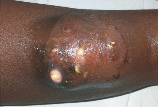
a proximal muscular weakness (LL>UL), periorbital pinkish discoloration
papular lesions on knuckles and elbow, and discharging whitish deposit
at both knee joints (Fig. 1). Investigations revealed
anemia (hemoglobin 8.4 gm/dL), transaminitis, C-reactive protein (72
mg/L), erythrocyte sedimentation rate (104 mm FHR), creatinine
phosphokinase (1012 U/L), and positive antinuclear antibody (ANA) test.
A diagnosis of juvenile dermatomyositis with calcinosis cutis was
considered. We administered methylprednisolone pulse therapy for 3 days,
followed by oral steroid, which led to marked symptomatic improvement.
 |
|
Fig. 1 Whitish lesions with deposits
and discharge in the region of left knee joint.
|
Calcinosis cutis is a pathological condition of
abnormal deposition of calcium in the skin and subcutaneous tissue. It
is classified into dystrophic, metastatic, idiopathic, iatrogenic and
calciphylaxis groups. Dystrophic is the most common type and usually
associated with connective tissue disorders. It is frequently
distributed at elbows and knees joints, and can lead to pain, chronic
ulceration, and secondary infection. Treatment includes calcium channel
blockers, colchicine, minocycline, warfarin, aluminum hydroxide,
bisphosphonates and probenecid.


