|
|
|
Indian Pediatr 2014;51: 243 |
 |
Bullous Impetigo
|
|
Shylaja Someshwar and HR Jerajani
Department of Dermatology, MGM Medical College, Navi
Mumbai, India.
Email: [email protected]
|
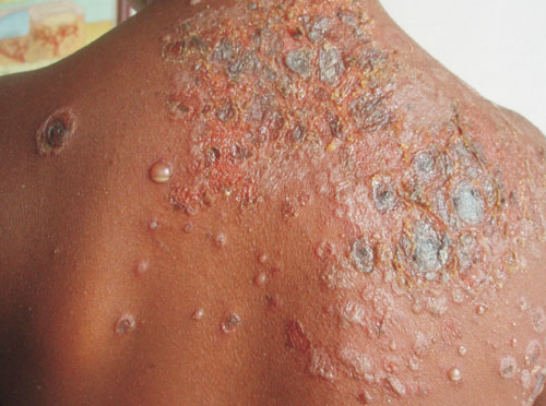
A 5-year-old girl presented to us with complaints of
exudative painful lesions on the face and upper back for 6
days. The lesions started as multiple fluid filled lesions
on the face which turned turbid, ruptured, spread and
involved the back. On examination, multiple pustules with
level of pus (positive hypopyon sign) were seen in few
lesions; erosions and honey coloured crusts were noted on
the upper back (Fig. 1) and face. Culture of
pus showed the growth of Styphylococcus aureus. She
was treated with a 7 day course of antibiotics following
which lesions cleared.
 |
|
Fig. 1 Crusted lesions with
bullae the arrow showing level of pus (Hypopyon
sign).
|
The lesions of bullous impetigo are
commonly seen on the face, trunk and extremeties which are
vesicles to begin with and later becoming pus filled,
followed by rupture and crusting. When the patient is in the
erect position, the pus that is heavier settles down giving
a positive hypopyon sign. Bullous impetigo has to be
differentiated from bullous erythema multiforme (typical
targetoid lesions), bullous lupus erythematosis (systemic
involvement), bullous pemphigoid (rare in childhood), and
subcorneal pustular dermatosis (sterile and classically
involves intertriginous areas).
|
|
|
 |
|
