|
|
|
Indian Pediatr 2012;49: 240-241
|
 |
Pemphigus Foliaceus
|
|
Ghalamkarpour Fariba, Azin Ayatollahi and Somayeh
Hejazi
From the Skin Research Center, Shahid Beheshti
University Shohada-e Tajrish Hospital, Shahrdari St, 1989934148, Tehran,
Iran.
Correspondence to: Ghalamkarpour Fariba, Skin Research
Center, Shahid Beheshti University (MC), Shohada-e Tajrish Hospital,
Tehran, Iran.
Email:
[email protected]
Received: August 30, 2010;
Initial review: September 23, 2010;
Accepted: Febuary 01, 2011.
|
Pemphigus foliaceus is an autoimmune blistering disease, which affects
the skin but rarely affects the mucosae. There are two variants of
pemphigus foliaceus : endemic and sporadic. Erythroderma due to
pemphigus foliaceus is unusual and its occurrence in a child is very
rare. We describe a case of erythrodermic pemphigus foliaceus in a 12-
year- old boy.
Key words: Foliaceus, Iran, Pemphigus.
|
|
Pemphigus foliaceus is an autoimmune
blistering disease, which affects the skin but rarely affects the
mucosae [1]. Erythroderma due to pemphigus foliaceus is unusual and
its occurrence in a child is very rare . We describe this entity in
a 12- year- old boy.
Case Report
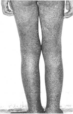
A 12-year-old boy presented to us with widespread
skin lesions of 4 months duration. Erythematous and crusted lesions
first appeared on his scalp and within a few days it became
generalized and then erythrodermic (Fig. 1). Scaling
and exudation were also seen. Different antibiotics and topical
steroids were prescribed without significant improvement. The child
also complained of hearing loss since one month. On physical
examination, erythroderma with severe scaling and malodorous
discharge was seen. There was mild palmoplantar keratoderma and
scales covered the entire scalp. There were two small vesicles along
the ulnar side of right palm. The mucosal surfaces and nails were
normal. He had two small non tender submandibular lymph nodes.
External auditory canal was filled with scales and crusts. Pinna was
tender on palpation. After removing the crusts, external auditory
canal was found to be red and swollen. Routine laboratory tests were
normal except erythrocyte sedimentation rate which was elevated
(52mm/hr). Giardia cyst was found in stool exam. KOH
examination from the scalp scales was negative for dermatophytes.
Lesional and perilesional biopsy were taken with impression of
pemphigus foliaceus, eczema, psoriasis, and erythroderma due to
dermatophytosis.
 |
|
Fig.1 Generalized crusted lesions
in Pemphigus foliaceus.
|
The microscopic examination showed a subcorneal
cleft in the granular layer. A few acantholytic cells were also
seen. Mixed inflammatory infiltrate with lymphocytic predominance
was seen in dermis. Direct immunofluorescence performed on
prelesional specimen showed deposits of C3 and IgG in the upper part
of the epidermis compatible with pemphigus foliaceus. Oral
prednisolone 30 mg/d and azathioprine 50 mg/d were started. Proper
treatment was instituted for external otitis and Giardia
cyst. Prednisolone was increased to 50 mg/d due to poor response to
treatment. A few days later the lesions began to improve. The child
was discharged after 45 days of admission. Follow-up was not
possible.
Discussion
Pemphigus foliaceus is an autoimmune disease that
is characterized by the presence of autoantibodies against the cell
surface of keratinocytes, which leads to destruction of epidermal
cell junctions. Blistering in this group of autoimmune disease
occurs in upper parts of the epidermis, either in the granular layer
or just beneath the stratum corneum. Pemphigus foliaceus comprises
of two major categories: (i) sporadic form; and (ii)
endemic pemphigus foliaceus also known as fogo selvagem (wild fire)
[1-4].
Fogo selvagem primarily affects children in
contrast to the sporadic form of pemphigus foliaceus which is
generally a disease of the middle aged and elderly people. It is
rare among children [2] with the average age at presentation being
7.7 years [5]. The youngest patient reported so far was a 18-
month-old child [5].Childhood pemphigus foliaceus appears to be
slightly more common in boys [1]. The most common sites of
involvement are scalp and face, followed by the trunk or upper
extremities [6]. Lesions of the groin or pubic area are rarely
reported. Mucous membranes are generally not involved. Pemphigus
foliaceus may remain localized or become generalized. Progression of
disease to multiple cutaneous sites occurrs in more than half of the
cases [1].The most common primary lesions appear on the face and the
scalp as superficial bullae which rapidly rupture and leave scales
and crusts. Sometimes only scales and crusts with erythematous bases
may be seen [6,9]. Generalized erythroderma has been reported in 2
cases of childhood pemphigus foliaceus [3]. Triggering factors such
as UV exposure, drugs and various infections have been proposed as
provoking factors for the disease [1]. Our patient suffered from
otitis and giardiasis which may have precipitated pemphigus
foliaceus. In most cases of pemphigus foliaceus, the diagnosis is
based on histopathology. Pemphigus foliaceus may initially be
misdiagnosed as impetigo, seborrheic dermatitis, eczema, psoriasis
and dermatitis herpetiformis. In majority of cases topical and
systemic corticosteroids are used for the treatment.
Contributors: All authors contributed to
literature search, review of literature, patient diagnosis and
managements and drafting the manuscript.
Funding: None; Competing interests:
None stated.
References
1. Metry DW, Hebert AA, Jordon RE. Non endemic
pemphigus foliaceus in children. J Am Acad Dermatol. 2003;49:187-9.
2. Amagai M. Pemphigus. In: Bolognia JL,
Jorizzo JL, Rapini RP, Horn TD, Mascaro JM, Mancini AJ, et al.,
editors. Dermatology. Edinburgh: Mosby. 2008. p.417-28.
3. Mehravaran M, Morvay M, Molnar K, Oláh J,
Korom I, Husz S, et al. Juvenile pemphigus foliaceus. Br J
Dermatol. 1998;139:496-9.
4. Diaz LA, Sampaio SA, Rivitti EA, Martins CR,
Cunha PR, Lombardi C, et al. Endemic pemphigus foliaceus (fogo
selvagem), I: clinical features and immunopathology. J Am Acad
Dermatol. 1989;20:657-9.
5. Kahn G, Lewis HM. True childhood pemphigus.
Pemphigus foliaceus in an 18-month-old child: immunofluorescence as
a diagnostic aid. Am J Dis Child. 1971;121:253-6.
6. Goodyear HM, Abrahamson EL, Harper JI.
Childhood pemphigus foliaceus. Clin Exp Dermatol. 1991;16:229-30.
7. Qureshi WA, Ali A, Bhol KC, Ahmed AR. Juvenile
pemphigus foliaceus treated with sublesional corticosteroids. Int J
Dermatol. 1997;36:848-50.
8. Rosella M, Masia IM, Satta R, Cottoni F.
Pemphigus foliaceus in a child. Pediatr Dermatol. 1996;13:259-60.
9. Sotiriou L, Herzenson S, Jordon RE. Childhood
pemphigus foliaceus: report of a case. Arch Dermatol.
1980;116:679-80.
|
|
|
 |
|
