|
Piebaldism is a autosomal
dominant disorder of localized amelanotic
patches as a result of permanent localized
absence of melanocytes. GSD1a is autosomal
recessive disorder of carbohydrate metabolism
characterized by deficiency of
Glucose-6-phosphatase. We herein report a rare
case of GSD type1a with piebaldism.
Case Report
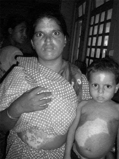
A 3½-year old male child was admitted with
history of gradual distention of abdomen since
one year of age. The child had localized
hypopigmented areas over anterior scalp, both
elbows, knees, over abdomen since birth, which
were similar to those in his mother and elder
sister (Fig.1). Second degree
consanguinity was present in parents. The
maternal grandparents also had 2nd degree
consanguinity. At around 9 months of age, the
child had an episode of convulsion on overnight
fasting. Three more convulsions were reported
later. Child had overwhelming hunger, poor
growth, frequent lethargy and difficult arousal
from overnight sleep. There was no history of
recurrent infections or muscle weakness. On
examination, the child had doll like face, fat
cheeks, short stature, relatively thin
extremities, protuberant abdomen, delayed
milestones. Deafness was absent. He was
underweight and stunted. There was no family
history of protuberant abdomen or hepatomegaly.
Liver span was 13 cm and firm. Spleen was not
palpable. Laboratory workup revealed haemoglobin
10g/dL, complete blood count including
differential count, platelet count, bleeding
time and coagulation profile were within normal
limits. Random blood glucose level was 46 mg/dL.
Liver enzymes and uric acid were within normal
limits. Serum cholesterol was 186 mg/dL (Normal
155 mg/dL), triglyceride was 320 mg/dL (Normal
56 mg/dL). Lactate-creatinine ratio was
estimated in urine which was 0.9 mmol/mmol. USG
revealed hepatic enlargement. Blood gas analysis
revealed metabolic acidosis with respiratory
compensation. EEG and MRI brain were within
normal limits. Percutaneous liver biopsy
revealed enlarged pale hepatocytes distended
with glycogen, compressing sinusoids and giving
a mosaic pattern. Intracellular glycogen was
demonstrated with periodic acid Schiff reaction,
readily digested by diastase. Slender periportal
fibrous band was present. Histological features
were consistent with liver glycogenosis. Genetic
sequencing was done for all exons of 17q21
gene. A G727T gene splice mutation was
diagnosed in exon 5 of 17q21 gene. The
child was homozgyous for the mutation. Genetic
analysis of parents for GSD1a could not be
arranged. Punch biopsy of piebald skin revealed
absence of melanocytes and melanin pigment in
white depigmented area by Manson Fontana stain.
The case was diagnosed as a case of Glycogen
storage disease 1(a) with piebaldism.
 |
|
Fig.1
Piebaldism seen in patient and mother.
|
After starting dietary
therapy, the fasting blood sugar increased to
80-85mg/dL, urinary lactate-creatinine ratio was
0.07 mmol/mmol. Annual USG surveillance and
sunprotective measures were advised. Skin
grafting for repigmentation of piebald skin is
also planned.
Discussion
GSD1(a) is an autosomal
recessive disease caused by mutations at loci
17q21 [1]. This patient have G727T splice
mutation which may be manifested due to second
degree consanguinity among parents and maternal
grand parents. G727T mutation is a common
reported mutation in Japanese and Chinese
patients [2]. In Indian children, this mutation
is relatively rare. In GSD1a cases, stringent
genotype phenotype relation is not found [3].
The piebald skin have mutation of KIT gene
inherited in a autosomal dominant manner. These
two mutations appears to be unrelated and a
chance finding. Histological features in liver
glycogenoses include documented fatty change,
nuclear hyperglycogenation and fibrosis [4]. We
could demonstrate glycogen in hepatocytes in our
patient with slender periportal fibrosis. Our
patient has responded to uncooked cornstarch
feeding to maintain the blood sugar level. Young
infants need continuous nasogastric feed with
sucrose free low lactose formula enriched with
maltodextrine for this purpose [5].
The differential diagnosis of
Piebaldism are Addison disease, albinism,
vitiligo, Vogt koyanagi Harada syndrome,
Waardenburg Syndrome, etc. The nonprogressive
nature of the hypopigmentation and absence of
associated features rules out the other
possibilities. Piebaldism is one of the
cutaneous signs of Waardenburg syndrome, along
with heterochromia of iris, lateral displacement
of inner canthi, and deafness [6]. The present
case did not have deafness or facial features of
Waardenburg syndrome. The KIT mutation in
vicinity of codon 20 of 4q12 chromosome leads to
the usual phenotype of static piebaldism [7].
The depigmented skin in piebaldism is
unresponsive to medical and light treatment.
Autologous punch grafting for repigmentation in
piebaldism may be considered [8].
Contributors: MB and RB
diagnosed the case. BG and NS prepared the
manuscript and managed the case.
Funding: None;
Competing interests: None stated.
References
1. Brody LC, Abel KJ,
Castilla LH, Couch FJ, McKinley DR, Yin G, et
al. Construction of a transcription map
surrounding the BRCA1 locus of human
chromosome17. Genomics. 1995; 25:238-47.
2. Okubo M, Aoyama Y,
Kishirmoto M, Shishiba Y, MuraseT.
Identification of point mutation (G727T) in the
glucose 6 phosphatase gene in Japanese patients
with glycogen storage disease type1a and carrier
screening in healthy volunteers. Clin Genetics.
1997;51:179-83.
3. Chou JY, Mansfield BC.
Mutations in the glucose 6 phosphatase-á (G6PC)
gene that cause type1a glycogen storage disease.
Hum Mutation. 2008;29:921-30.
4. Hasan Ö. Glycogen storage
diseases: New perspectives. World J
Gastroenterol. 2007; 13:2541-53.
5. Rake JN, Visser G, Labrune
P, Leonard VJ, Ullrich K, Smit GP. Guidelines
for management of glycogen storage disease type
1-European Study on Glycogen Storage Disease
type 1(ESGSD-1). Eur J Pediatr. 2002;161:112-9.
6. Jan JA, Stroedter L, Haq
AU. Association of Shah Waardenburg syndrome: a
review of 6 cases. J Pediatr Surg.
2008;43:744-7.
7. Ward KA, Moss C, Sanders
DS. Human piebaldism: relationship between
phenotype and site of kit gene mutation. Br J
Dermatol. 1995;132:929-35.
8. Garg T, Khaitan BK,
Manchanda Y. Autologous punch grafting for
repigmentation in piebaldism. J Dermatol.
2003;30:849-50.
|


