|
|
|
Indian Pediatr 2011;48: 239-241 |
 |
Esophageal Diverticulum Secondary to Impacted
Foreign Body |
|
Rekha Harish, Ashu Jamwal, Gurjeet Singh* and Arvind Kohli*
From
the Departments of Pediatrics and Cardiothoracic Surgery*, Government
Medical College, Jammu.
Correspondence to: Dr Rekha Harish, 11-B, Shastrinagar
Extn, Near Dogra Academy, Jammu, J&K State 180004, India.
Email: [email protected]
Received: June 3, 2009;
Initial review: September 4, 2009;
Accepted: November 30, 2009.
|
We report a two year old child who developed a large esophageal
diverticulum over a period of ten months following ingestion of a
multispiked leaf of Quercus semicarpipholia. Though the
endoscopic removal of foreign body was successful, it did not relieve
the symptoms and patient required surgical resection of the diverticulum.
Patient is asymptomatic after 4 months of follow up.
Key words: Child, Diverticulum, Esophagus, Foreign body.
|
|
Foreign body ingestion is frequent in
children, especially between six months to three years of age owing to
their inherently inquisitive nature [1]. Though majority of ingested
foreign bodies traverse the gastrointestinal tract without any adverse
effects, occasionally they can get impacted resulting in various
complications [2]. A two years old child is reported with an impacted
woody tree leaf in esophagus, producing a valve effect causing partial
obstruction and development of a large, secondary esophageal diverticulum
over a ten months period.
Case Report
A two year old male child was brought with history of
persistent vomiting following any solid food ingestion and progressive
weight loss for the last ten months. The child had a normal growth and
development till fourteen months of age when he had sudden choking with
cough while playing. The initial two vomitings contained small amounts of
fresh blood but later it contained only the ingested solid food. The
vomitings persisted despite several medications and gradually the mother
noticed that the child tolerated small frequent fluid feeds which
comprised mainly of water and milk.
Examination revealed an afebrile, pale and malnourished
child (PEM grade II). Systemic examination and the biochemical laboratory
work up was within normal limits. Chest radiographs did not reveal any
abnormality. An upper gastrointestinal obstruction was suspected and
the child was subjected to endoscopy.
A vegetative foreign body in the form of two pieces of
semilunar thick tree leaves was observed blocking the lumen of esophagus
with suspicion of diverticula proximal to it. The foreign body removed
endoscopically was a single leaf 3cm × 2cm with thorny peripheral edges (Fig
1), which had caused impaction. The leaf was torn in the middle
with two pieces acting as valvular flaps and allowing fluids to trickle
down. It was identified as leaf of Quercus semicarpipholia, a
species commonly found in hilly areas of J & K state. However,
endoscopic removal did not relieve the symptoms and a barium esophagogram
(Fig 2) done revealed a large diverticulum at the
midesophagus level with dilated proximal portion of esophagus.
Computerised tomography chest confirmed these findings. Patient had an
episode of chest infection which responded to antibiotics. He was then
transferred to Cardiothoracic Unit for surgery. Intraoperatively there was
a big diverticulum in relation to the mid esophagus which was excised and
end to end anastomosis was done. Patient is symptom free after four
months follow up.
 |
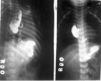 |
Fig. 1 Quercus leaf with spiny edges which was removed by
endoscopy.
|
Fig
2 Barium swallow showing the esophageal diverticulum as a
large outpouching from the right lateral wall of the esophagus. |
Discussion
Early recognition and treatment of the esophageal
foreign bodies is imperative because complications can be serious and life
threatening viz perforation, extraluminal migration, mediastinitis,
hemorrhage, aorto-esophageal fistula, stricture and esophageal
diverticulum [3]. Most of the
esophageal diverticula occur in middle aged adults and elderly people,
however rarely they may occur in children [4]. Macpherson, et al.
[3] in a study of esophageal foreign bodies in 118 children
reported diverticulum in one case. Patients may remain asymptomatic
or may present with dysphagia, regurgitation, halitosis or aspiration
pneumonia. Retention of undigested food in large diverticula results in
regurgitation, nocturnal cough and aspiration pneumonia [4].
Diagnostic modalities include barium swallow, upper GI
endoscopy, and computed tomography. Barium radiography is generally the
procedure of choice. In addition to being excellent at defining the
structural appearance of diverticula, barium swal-low may also provide
clues to underlying motility disorders that may be involved in
diverticular formation. On CT scan, large diverticula of esophagus may
manifest as air and/or fluid filled structures communicating with the
esophagus [5]. Endoscopy can be performed to rule out underlying
structural lesions.
Asymptomatic and minimally symptomatic esophageal body
diverticula do not require treatment. Surgical management described for
symptomatic mid thoracic or epiphrenic diverticula are extended myotomy
and diverticulectomy with an anti reflux procedure. An abdominal
laproscopic approach may be feasible for some patients with epiphrenic
diverticula [6]. Endoscopic
treatment of giant mid-esophageal diverticula has been occasionally
reported [7].
There are a very few case reports of esophageal
diverticula in children following impacted foreign body. Akhter, et al.
[8] reported a two and half year old boy who developed a
large esophageal diverticulum following an impacted plastic button which
remained undiagnosed for 18 months. Herman, et al. [9] reported two
pediatric patients of 7 and 2 years, who presented with progressive
dysphagia of 4 and 6 months period, respectively due to esophageal
stictures and secondary diverticulum due to unrecognised impacted foreign
bodies [9]. The present case had
developed a large mid esophageal pulsion diverticulum as a result of
impacted tree leaf for a prolonged period of ten months. The leaf
was woody and had multiple small spikes on the margins which lead to
circumferential impaction. The breech in the middle allowed the patient to
sustain life on fluids alone for ten months.
Contributors: RH and AJ were responsible for
diagnosis, investigative workup, conservative management, compiling
literature and preparing the manuscript. GS and AK were involved in
surgical management and assisted in drafting.
Funding : None.
Competing interest: None stated.
References
1. Little DC, Shah SR, St Peter SD, Calkin CM, Morrow
SE . Esophageal foreign bodies in the pediatric population:
Our first 500 cases. J Pediatr Surg. 2006;41:914-8.
2. McGahren ED. Esophageal foreign bodies. Pediatr Rev.
1999;20:129-33.
3. Macpherson RI, Hill JG, Otherson HB, Tagge EP, Smith
CD. Esophageal foreign bodies in children: diagnosis, treatment and
complications. Am J Radiol. 1996;166:919-24.
4. Nichols FC. Diverticula of the esophagus. Surg Clin
North Am. 2005;85:495-503.
5. Kim KW, Berkmen YM, Auh YH, Kazam E. Diagnosis of
epiphrenic esophageal diverticulum by computed tomography. Comp Tomogr.
1988;12:25-8.
6. Fernando HC, Luketech JD, Samphire J, Alveo RM,
Christina NA, Buenanleira PO. Minimally invasive operation for esophageal
diverticula. Ann Thorac Surg. 2005;80:2076-80.
7. Nishimoto Y, Taguchi T, Ogita K, Hashizume M, Suita
S. Endoscopic diverticulectomy for symptomatic pediatric esphageal
diverticula. Pediatr Surg Int. 2005;21:50-3.
8. Nawaz A, Jacobsz A, Herticant J, Salem AH. An
unusual presentation of a retained esophageal foreign body. Ann Saudi Med.
1998;18:164-6.
9. Herman TE, McAlistar WH. Esophageal diverticula in
childhood associated with stictures from unsuspected foreign bodies of the
esophagus. Pediatr Radiol. 1991;21:410-2.
|
|
|
 |
|
