|
|
Images Indian Pediatrics 2008; 45:221 |
|||
|
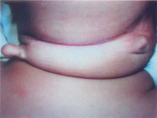
Bilateral Cervical Chondrocutaneous Branchial Remnants |
|||
|
A 4-month-old boy presented with bilateral neck lesions noticed since birth. Both lesions were located in the lateral neck anterior to sternocleidomastoid muscle and had firm but elastic texture (Fig. 1). Right was in the lower middle third of neck, was pediculated and measured 1.5 cms while left was sessile, measured 1.0 cm and located in upper middle third of neck. The overlying skin was similar to surrounding skin of neck and there was no discharge, tenderness or any other sign of inflammation. A thorough physical examination was unremarkable. Ultrasonography of genito-urinary system revealed no abnormality. Surgical excision was done under general anesthesia. The cartilaginous part was found attached to aponeurosis of sternocleidomastoid and sheath of external jugular vein on either side. No connection with deeper structures was noted. Histopathological examination showed polypoidal tissue lined by normal epidermis. Deeper dermis contained mature fat and well circumscribed lobules of mature hyaline cartilage.
Cervical chondrocutaneous branchial remnants are uncommon benign congenital lesions found in lateral neck and appear similar to "accessory tragi". Bilateral lesions are very rare. Previously described under a variety of names including tags, wattles, appendages, polyps, vestiges, accessory auricles or tragi, rests, choristomas, hamartomata, papillomata, fibromata etc., the clearer designation was proposed in 1997 and has been followed thereafter. These lesions are more often seen in males and originate from second branchial arch. Surgical removal is simple and usually undertaken for cosmetic reasons. A high incidence of associated anomalies mandates a meticulous physical examination and ultrasonogram of genitourinary tract. Goldenhar, Treacher-Collins and some other well characterized syndromes may include cervical or preauricular remnants. The clinical appearance and presence of central cartilage core eliminates the possibility of any other differential diagnosis for the cervical lesions. Devidayal,
|
![]()