|
|
Case Reports Indian Pediatrics 2006; 43:255-257 |
||||
|
Peripheral Gangrene: An Uncommon Manifestation of Disseminated Tuberculosis |
||||
|
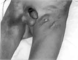
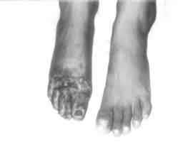
Tuberculosis is a very common condition in India with varied presentations. Peripheral ‘gangrene as a manifestation of tuberculosis is very uncommon. We present a child with this uncommon manifestation of a common disease. Case Report An 8-year-old male child presented with a non-healing ulcer over the sole of left foot following a puncture wound sustained while playing in the fields six months back. A swelling appeared over the left groin 2-3 weeks later, which subsequently invaded the overlying skin and broke down leaving a non- healing ulcer. He complained of cough with fever for past 20 days with no sputum, hemoptysis, or chest pain. Four days back, he developed sudden severe pain over the right fore foot followed by bluish discoloration, swelling and inability to move the foot and toes. There was no history of any transient ischemic attack, abdominal pain, hematuria, Raynaud phenomenon, malena or claudication. There were no bleed from any sites, neurological or visual symptoms or any joint pains or swellings. He had been well prior to this illness and was not on any medication. There was no family history or any known contact with tuberculosis. On examination his weight for age was 60%. There was no pallor, rash, petechiae, livedo reticularis, and joint swellings. All peripheral pulses including femorals, popliteals, posterior tibials, dorsalis pedis of both the limbs were normally palpable. Blood pressure in right upper limb was 110/50 mmHg; right lower limb 112/84 mmHg, left upper limb 112/54 mmHg, leftlower limb 120/81 mmHg. No bruits were heard. Lymph nodes were palpable in the left inguinal region (the largest measuring 3 cm × 1.5 cm). The ulcer overlying the lymph node measured 2 cm × 2 cm (Fig. 1). His right foot was gangreneous with no movement of the toes (Fig. 2). The wound over the left sole was a deep ulcerated one with a thick overhanging margin and no discharge. Chest examination showed decreased air entry on the left side of the chest. Cardiovascular system, per abdomen and central nervous system examination including sensory examination was normal.
On investigations his hemoglobin was 12 g/dL, TLC 5600 with 68% polymorphs and platelet count was 1.8 lacs. PT (14s against a control of 14s) and PTTK (31s against control of 32s) were normal. ESR was 90-mm/1st hour; CRP was negative. Blood ELISA for tuberculosis 1.46 (>1.11 is considered positive). Mantoux with 5 TU PPD was 8 mm. Random blood sugar was 90 mg/dL, S. urea 21 mg/dL, S. creatnine 0.8 mg/dL. Urine microscopy (centrifuged sample) showed no hematuria. Chest X-ray showed homogenous opacity on the left side of chest, which on ultrasonography was confirmed to be a loculated pleural effusion. Pleural fluid was straw with 1000 lymphocytes/mm3, sugar 30 mg/dL and proteins 8 g/dL. Adenosine deaminase was positive in the pleural fluid 123.5 U/L (>60 u/L is considered as positive). Both pleural fluid and sputum were negative for AFB.
Doppler study of the right lower limb revealed patent common femorals, superficial femorals, popliteals, anterior and posteriors tibials, dorsalis pedis with normal blood flow, good spontaneous color with triphasic wave-form pattern and normal velocity. The corresponding veins were also patent, compressible and showed spontaneous color fills in. FNAC of inguinal lymph node revealed casseous material with no granulomas. Skin biopsy from site of scrofloderma showed a sinus tract with focal ulceration of overlying epidermis. The tract was lined by chronic inflammatory infiltrate with a few epithelial cell granulomas and Langhans giant cells. Stain for AFB and fungal hyphae were negative. Skin biopsy from site of non-healing ulcer was also suggestive of chronic inflammation. Investigations for vasculitis syndromes were planned but could not be pursued due to financial constraints, however the patient did not fit into any other vasculitis syndrome clinically. In view of long history, casseous necrosis of lymph nodes and chronic inflammation on histopathology, the child was started on four drugs autitubercular therapy (ATT) along with short course of antibiotics (ceftazidime and cloxacillin). He showed steady improvement with the gangrenous foot drying up and a definite line of demarcation forming distal to the initial one. He was then referred to the orthopedic surgeon for amputation. The post operative course was smooth with complete wound healing. Repeat chest X-ray six months later showed complete clearance. Both the ulcers also showed good healing in response to ATT in 2 months. Discussion The mode of onset and progression of symptoms in this child suggests that he acquired tubercular infection as primary inoculation into the wound on his left foot. Playing barefoot on ground contaminated with infectious sputum can infect children in this manner. The site of predilection for inoculation of tuberculosis is the lower extremity, as this is most likely to be traumatized. Having traveled to the left inguinal lymph nodes the organisms infected the overlying skin to cause another manifestation of cutaneous tuberculosis, scrofloderma. In most reports this is the commonest form of cutaneous tuberculosis seen. In a study done at Chandigarh, which analyzed childhood cutaneous tuberculosis over 25 years, scrofloderma accounted for 53.3% of all cases(1). Spread of the infection to the lungs then followed causing systemic symptoms. Gangrene resulting from tuberculosis is uncommon. Itin, et al.(2) described symmetrical peripheral gangrene in a patient with disseminated tuberculosis. As mycobacterium tuberculosis was isolated from the blood culture, it was thought that symmetrical peripheral gangrene resulted from embolization of arterioles by the tubercular bacilli. Lopez, et al. reviewed six cases of tubercular pulmonary gangrene with AFB positive sputum smears. Four patients died and two responded to ATT(3). Our patient presented with disseminated tuberculosis and peripheral gangrene for which no cause could be ascertained clinically. Vasculitis is a known association with tuberculosis. Gangrene secondary to tuberculosis has been reported at various sites of the body like scrotum, penis, small intestines and retina(4-6). Many of the manifestations seen in tubercular meningitis are also due to vasculitis of the intracranial blood vessels. Our case did not fit into any other vasculitis syndrome. The child showed complete recovery on ATT. The purpose of this case report is to highlight this uncommon manifestation of disseminated tuberculosis, which is a common condition in our country. Funding: None. Competing interests: None. | ||||
|
References | ||||
|
|
![]()