|
|
Letters to the Editor Indian Pediatrics 2003; 40:276-277 |
||||
|
Radiographic Manifestations of Acute Lymphoblastic Leukemia |
||||
|
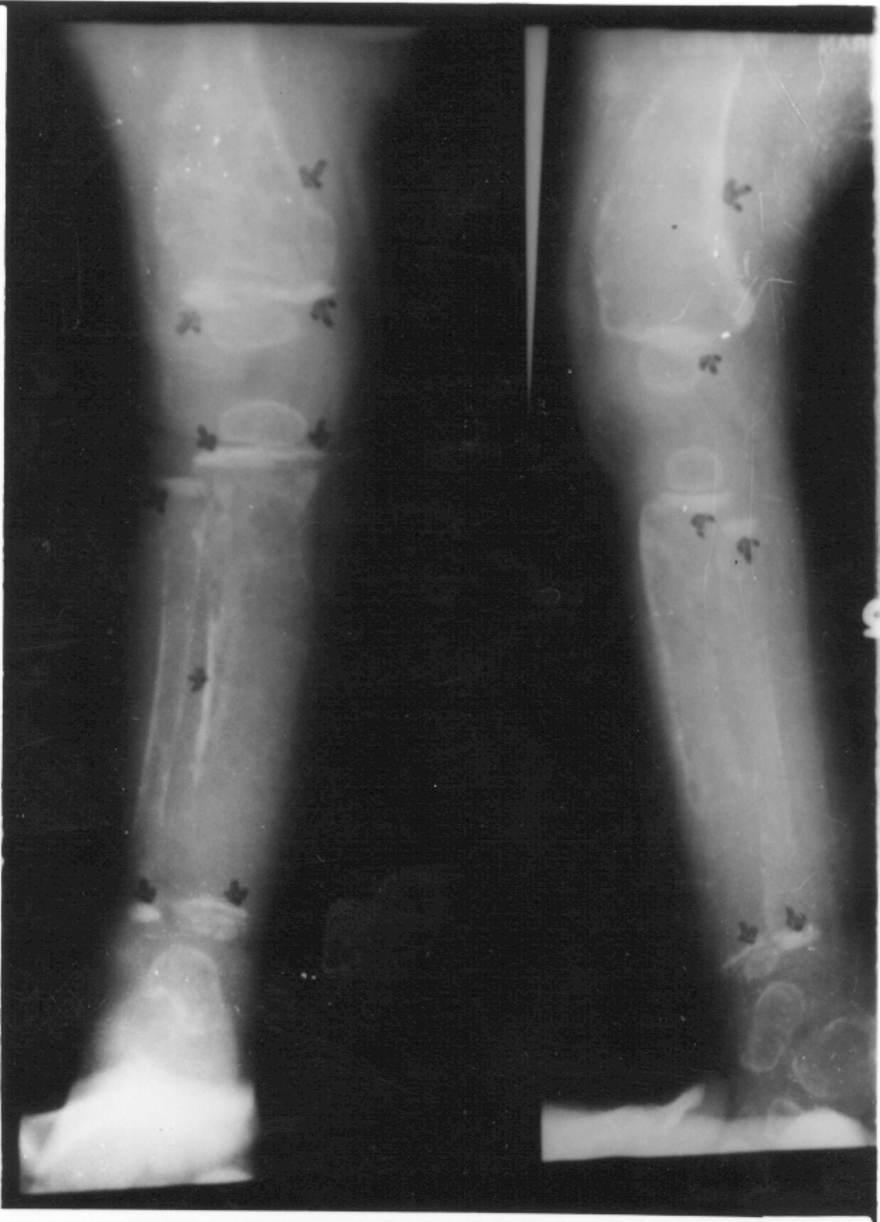
P, a 1-year-old child, presented with history of intermittent fever for 4 months, periorbital edema for 3 months and inability to sit or stand for 2 months. There was no history of hematuria, oliguria or polyuria. On examination, this child was found to be pale, with hepatomegaly of 4 cm below the right costal margin. Spleen was not palpable. Investigations at this point of time revealed hemoglobin-4.7 g/dL, TLC-22600/mm3, DLC-N13, L82, B2, platelet count - 85000/mm3. Peripheral smear showed normocytic, hypochromic blood picture with microcytes, leucocytosis and lymphocytosis. X-ray taken is shown in Fig 1. A provisional diagnosis of renal osteodystrophy/cystic disease of bone was made. However it was difficult to explain anemia and thrombocytopenia. An orthopedic consultation was sought and various causes for cystic lesions in the bone were thought of, but none could satisfactorily explain the clinical findings. A repeat peripheral smear examination revealed leucoerythroblastic blood picture with thrombocytopenia, includ-ing two blasts in the differential count. In view of this, a bone marrow was done which showed features suggestive of acute lymphatic leukemia (ALL).
Children with acute lymphoblastic leukemia can present with localized or diffuse bone pain, limp or failure to use an extremity(2-4). Bone pain is seen in 27-33% of children as the presenting complaint(3). Migratory joint pain with swelling and tenderness is often misdiagnosed as juvenile rheumatoid arthritis (JRA) or rheumatic fever(1,4). Sometimes the presentation can mimic that of osteomyelitis or septic arthritis. Jonson et al.(2) reviewed the records of 296 children with ALL to determine the relation-ship between bone pain and the hematological findings at diagnosis. They found that children in the group with musculoskeletal symptoms that over-shadowed the hemato-logic manifestations of leukemia, 46% of children had normal total leucocyte count and platelet count, in addition to 33% of them having no circulating blast cells. Our index case initially had anemia and thrombo-cytopenia. Later repeat examination revealed 2 circulating blast cells. However, anemia was significant, requiring blood transfusions. Radiographic abnormalities in patients with leukemia range between 47 and 70%(3,4). The common abnormalities des-cribed are geographic osteolysis in 38% (11-90%), metaphyseal bands of increased and/or decreased density in 34% (7.5-88%), diffuse osteoporosis in 27% (16-41%), periosteal reaction in 18% (2-50%), mixed osteolysis and osteosclerosis in 18%, osteosclerosis without lytic defects in 6% and permeative bone destruction in some cases(5). These changes are most easily seen in the long bones, especially around the areas of rapid growth (e.g., the knees, wrists and ankles). There are abnormalities in mineralization and bone metabolism(4). Our index case had generalized osteoporosis causing moth eaten appearance, metaphyseal bands of increased and decreased density and periosteal reaction. Heinrich et al.(5) reported that presence of 5 or more skeletal lesions at diagnosis is asso-ciated with a delay in diagnosis (symptomatic for 54 days) and decreased survival (76%) when compared to children with 1-4 lesions who were diagnosed early (symptomatic for 22 days) and had 100% survival. Our child was started on chemothrapy and the first check bone marrow had normalized without evidence of leukemia. Subsequently, the child was lost to follow up. I.E.D’Souza,
|
![]()