1.gif (1892 bytes)

Images in Clinical Practice

Indian Pediatrics 2002; 39:308  

Congenital Neuroblastoma with Cutaneous Metastases


A six-day boy was admitted to hospital with hyperbilirubinemia. He was born by normal vaginal delivery in a local hospital at 40 weeks’ gestation and weighed 3450 g. The pulse rate was 180/min, respiratory rate was 44/min and blood pressure was 64/42 mmHg. The abdomen was distended. On physical examination, two subcutaneous nodules on the abdominal wall and a lower quadrant abdominal mass were felt. Shortly after admission these firm, blue-gray nodules distributed over his body (Fig. 1). A biopsy was performed on a nodule on his right hip, and the histopathological picture confirmed the diagnosis of neuroblastoma. The patient died 40 days after birth.

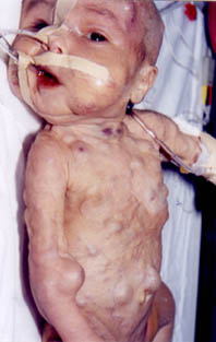
Neonatal tumors are rare. They comprise 2% of all pediatric malignancies, with an incidence of 1.58 to 3.65 per 100 000 live births. A neonatal unit with 3000 deliveries a year might expect to see a case once every 10 to 20 years. The most common neonatal tumor is neuro-blastoma, accounting for 28-39% of tumors in this period.

Neuroblastoma originates from neural crest cells of the adrenal medulla or sympathetic ganglia. The neoplasm can metastatsise to the liver, lymph nodes, bone marrow and skin in the neonatal period. In these infants, the first clinical manifestations usually result from the complica-tions of metastatic disease rather than the primary tumor.

Fig. 1. Subcutaneous metastases in a newborn infant with neuroblastoma.

Tamer Gunes,
Mustafa Akcakus,

University of Erciyes, Faculty of Medicine,
Department of Pediatrics,
Division of Neonatalogy,
38039, Kayseri, Turkey

E-mail:
[email protected]

Home

Past Issue

About IP

About IAP

Feedback

Links

 Author Info.

  Subscription