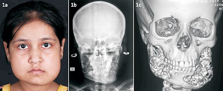
A 10-year-old girl presented with bilateral jaw swelling
noticed by her parents when she was 3 years old. Her father had
a similar history with his jaw swelling spontaneously regressing
by the 25 birthday. Examination revealed fullness of cheeks and
jaws with upward tilting of eyes exposing the sclera below
inferior limbus, giving, the so-called ‘cherubic’ appearance
(Fig. 1a). Radiograph revealed multiple thin-walled cystic
lesions involving rami and body of mandible (Fig. 1b),
reconfirmed on computed tomography images (Fig. 1c). Biochemical
investigations revealed mildly elevated total alkaline
phosphatase. She was diagnosed as familial cherubism and
reassured about the self-limiting disease course.
 |
| Fig. 1
Cherubism characterized by (a) Bilateral fullness of the
cheeks and jaws with slight upward tilting of the eyes,
giving a ‘cherubic’ appearance; (b) Radiograph of the
face showing multiple thin-walled cystic lesions
involving rami and body of mandible; (c) Computed
tomography of the face with 3D reconstructed images
showing multiple expansile soft-tissue lesions involving
both halves of the mandible causing areas of thinning
and destruction of the overlying bony cortex. |
Cherubism is a fibro-osseous disorder characterized by the
appearance of multilocular, expansile radiolucent lesions
involving mandible or maxilla that usually appear at 2-7 years
of age. Gain-of-function mutations in SH3BP2 gene have been
implicated. Differential diagnoses include brown tumor of
hyperparathyroidism, Noonan/multiple giant cell lesion syndrome,
fibrous dysplasia (as part of McCune-Albright syndrome), and
ossifying fibromas seen in hyperparathyroidism-jaw tumor
syndrome. Cherubism is self-limiting and begins to regress
spontaneously around the age of puberty.

