A 5-year-old girl, diagnosed with multi-system langerhans
cell histiocytosis (bone, liver, bone marrow and pituitary
involvement), was started on induction chemotherapy (weekly
cycles of vinblastine/prednisolone for 12 weeks). Two months
later, she developed progressive bluish-black discolouration
over the nose and fingertips along with darkening of the nail
beds without any itching, pain, redness, numbness, trauma or sun
exposure.
Several differentials were considered, Chikungunya fever is well
reported for causing an acute, brownish-black, centrofacial
hyperpigmentation [1]. It can persist for three to six months
after resolution of infection [1]. However, our child had no
fever, arthralgia or cytopenias to suggest an infectious
aetiology. Addisonian hyperpigmentation, which commonly involves
mucous membranes, flexures, palmar and plantar creases, the
areola, genitalia and pressure points (elbows and knees), was
ruled out clinically, as well as by the absence of hyponatremia,
hyperkalemia and acidosis [1]. Hyperpigmentation in thyroid
disorders also has a similar distribution as in Addison disease
[2]; however, the thyroid function test was normal. Exogenous
ochronosis, secondary to topical hydroquinone use, responsible
for bluish-black hyperpigmentation of the sun-exposed areas of
the face, was also ruled out as there was no history of such
application; neither was henna applied locally [1,2].
She had no preceding redness, scaling, pain, injury or
cutaneous eruptions to suggest post-inflammatory
hyperpigmentation [3]. Acanthosis nigricans, although
classically noted over the nape of the neck, axilla and groin,
can also develop over the face [4]. However, our child had
neither hyperglycemia nor obesity and the lesion in question
lacked the characteristic velvety thickening of acanthosis
nigricans [3]. The involvement of distal phalanges,
interphalangeal joints and oral mucosa, characteristic of
Vitamin B12 deficiency,
was absent in our child [2]. Moreover, her red blood cell
indices were normocytic and normochromic, ruling out this
possibility. Drug-induced acral hyperpigmentation was considered
after ruling out other differentials and vinblastine was
discontinued, following which the hyperpigmentation faded over a
period of 3 weeks, but did not disappear completely.
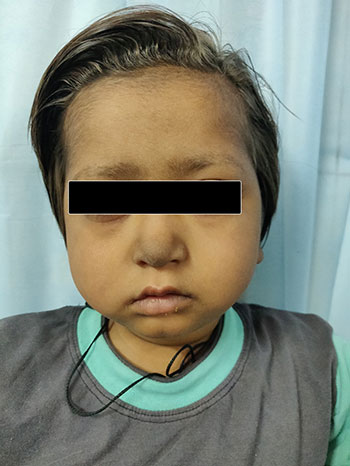 |
| Fig.1 |
Vinca alkaloids are notorious for causing extra-vasation injury
[4]. Supravenous hyperpigmentation has been reported with the
ABVD regimen which includes vinblastine (however, the causative
drug was not implicated) [5]. Vinorelbine, a vinca alkaloid, in
high doses is known to cause acral erythema [6].
Although drug-induced hyperpigmentation is responsible
for 10-20% cases of acquired hyperpigmentation [2], it has not
been reported with either vinblastine or prednisolone. Possible
mechanisms of drug induced acral hyperpigmentation include
increased melanin synthesis (secondary to cytotoxic effect on
melanocytes), cutaneous drug accumulation or iron deposits
following dermal vascular damage, and increased blood flow to
acral areas causes drug deposition [4,5].
Contributors:
AC: collected and analyzed data, drafted the paper; PKS:
conceptualized, analysed data, drafted and critically appraised
the manuscript.
Funding:
None; Competing interest: None stated.
REFERENCES
1. Khanna N, Rasool S.
Facial melanoses: Indian perspective. Indian J Dermatol Venereol
Leprol. 2011;77:552-64.
2. Bhalla M, Garg S. Acral
melanosis. Pigment Int. 2018;5:14-27.
3. Vashi AN, Kundu RV.
Facial hyperpigmentation: Causes and treatment. Br J Dermatol.
2013;169:41-56.
4. Payne AS, James WD,
Weiss RB. Dermatologic toxicity of chemotherapeutic agents. Sem
Oncol. 2006;33:86-97.
5. Pavey RA, Kambil SM,
Bhat RM. Dermatological adverse reactions to cancer
chemotherapy. Indian J Dermatol Venereol Leprol. 2015;81:434.
6. Heidary N, Naik H,
Burgin S. Chemotherapeutic agents and the skin: An update. J Am
Acad Dermatol. 2008;58:545-70.


