|
|
|
Indian Pediatr 2014;51: 510-511 |
 |
Ecthyma
|
|
Sidharth Sonthalia, *Archana Singal and
Rashmi Khurana
Kalyani-Escorts Hospital, Gurgaon, India and
*Department of Dermatology,
UCMS and GTB Hospital, Delhi, India.
Email:
[email protected]
|
An 8-year-old boy presented with a
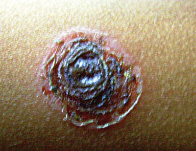
painful crusted lesion over the left forearm. Cutaneous
examination revealed a solitary coin-sized, indurated,
ulcerated, tender plaque with central brownish adherent
crust and yellowish-brown dried exudates at the margin (Fig.
1). There was no preceding history of any insect or
arthropod bite. The Gram stain from pus obtained from
underneath the crust revealed gram-positive cocci, and
culture grew both Group A
b-hemolytic
streptococci and Staphylococcus aureus. A diagnosis
of ecthyma was made; oral cefixime and topical mupirocin
ointment were prescribed along with removal of crust using
diluted white vinegar soaks. Complete healing with scarring
occurred within 2 weeks.
 |
|
Fig. 1 Solitary ulcer with central
adherent crust and yellowish-brown dried up exudates
at the ulcer’s margin.
|
Ecthyma denotes cutaneous bacterial
infection that extends deep into the dermis and heals with
scarring. It usually develops over disrupted skin on
extremities and rapidly develops into a vesicopustule and
finally a hemorrhagic crust. Differential diagnoses include
arthropod bites, leishmaniasis, ecthyma gangrenosum (vide
infra), pyoderma gangrenosum, Mycobacterium marinum
infection, and papulonecrotic tuberculid. Two related terms
need to be differentiated from ecthyma: Ecthyma
gangrenosum (a gangrenous ulcer with a central eschar
surrounded by an erythematous halo) a pseudomonal infection
that occurs in immuno-suppressed or gravely ill patients,
and Ecthyma contagiosum (solitary pustular lesions on
hands) resulting from the direct contact of damaged skin
with animal infected by a virus of Parapoxvirus group.
|
|
|
 |
|

