Naxos disease is a recessive form of
arrhythmogenic right ventricular dysplasia/cardiomyopathy
associated with a cutaneous phenotype characterized by
palmoplantar keratosis and woolly hair. It is caused by
mutations of the genes encoding desmosomal proteins [1]. Cardiac
disease has 100% penetrance by adolescence, manifested as
symptomatic arrhythmias, heart failure and sudden death. The
variant, Carvajal syndrome is characterized by younger age at
presen-tation and more pronounced left ventricular involvement.
Case Report
An 11-year-old girl of Indian origin born out
of 3
rd degree
consanguineous marriage presented with 2 transient epi-sodes of
syncope during exertion within a period of three months. Both
the times she recovered spontaneously before reaching the
hospital. She was previously asymptomatic except for thickening
of palmar and plantar skin with fissures noticed since early
childhood. None of her family members had a similar illness.
General examination revealed woolly hair and hyperkeratosis of
palms and soles. In the palms, hyperkeratosis was most marked in
the subungual areas. There were fissures on the plantar aspect
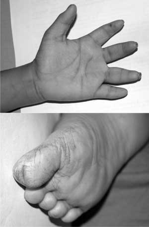
of big toe. In addition, distal phalanges of hands appeared
shorter than normal (Fig. 1). Rest of the general
examination including anthropometry was normal. Clinical
examination of cardiovascular system revealed a resting pulse
rate of 80/minute with normal volume and character of the
peripheral pulses. Blood pressure was 96/44 mm Hg in the left
upper limb in the sitting position. Cardiac apex was in the 5th
left intercostal space in the midclavicular line with normal
character, indicating cardiomegaly. First and second heart
sounds were normal and there were no additional sounds or
murmur.
 |
|
Fig. 1 Palmoplantar keratosis.
Note the subungual keratosis in the hands and fissures
in the sole. Short distal phalanges of fingers are also
obvious.
|
She was evaluated further to find out the
etiology of syncope. Baseline electrocardiogram (ECG) showed
complete right bundle branch block with left posterior hemiblock
indicating advanced myocardial disease. Transthoracic
echocardiogram showed mild ventricular dysfunction. Left
ventricle (LV) was predominantly involved with significant
chamber dilatation and the ejection fraction was 45%. ECG
obtained during subsequent episode showed ventricular
tachycardia (VT) at a rate of 150/ minute (regular wide QRS
tachycardia with north-west axis and deep S in V5 and V6)
suggesting VT as the etiology of syncope. Magnetic resonance
imaging did not reveal any fat deposits in the myocardium.
The phenotypical features and cardiac
manifestations along with history of consanguinity are
suggestive of arrhythmogenic cardiomyopathy with autosomal
recessive inheritance. These features are consistent with Naxos
disease, probably Carvajal variant. Family was counseled
regarding the disease and poor prognosis. She is being managed
with amiodarone and antifailure medications including carvedilol.
Two years from the initial diagnosis, her disease continues to
worsen with recurrent refractory episodes of ventricular
tachycardia and progressive cardiac failure.
Discussion
Naxos disease was first reported in 1986 by
Protonotarios, et al. [1] in patients from the Greek
island of Naxos. Apart from Naxos, cases have also been reported
from Italy, Turkey, Israel, Saudi Arabia and India. The variant
with more pronounced left ventricular involvement and clinical
overlap with dilated cardiomyopathy has been described in
families from Ecuador (Carvajal syndrome) [2].
Genetic studies have located two causative
genes, encoding for the desmosomal proteins plakoglobin and
desmoplakin. Homozygous mutation of the plakoglobin
gene truncating the C terminal of the protein causes Naxos
disease which maps to 17q21 [3]. Homozygous mutations of another
desmosomal component, desmoplakin which truncates the C terminal
of the protein and maps to 6p24 is identified in involved
patients from Ecuador [4]. The disease pathogenesis is linked to
the specific tissue characteristics of cardiac muscle. Cardiac
muscle consists of single myocytes connected by complex
intercellular contact sites called intercalated discs. Three
different types of intercellular junctions are located in
intercalated discs, namely adherence junctions, gap junctions
and desmosomes. Adherence junctions and desmosomes secure
mechanical coupling enabling synergistic contraction while gap
junctions serve electrical coupling allowing rapid spread of
action potentials. Plakoglobin is a common component of both
adherence junctions and desmosomes. At the adherence junctions,
it is connected to the actin cytoskeleton and at desmosomes to
the intermediate filaments of desmin. Desmoplakin is another
desmosomal protein that interlinks plakoglobin or plakophilin
with desmin intermediate filaments. Defects in linking sites (C
terminal) of these proteins interrupts cell to cell adhesion,
particularly under conditions of increased mechanical stress
leading to cell isolation and death. The result is progressive
loss of myocardium and fibro-fatty replacement. Surviving
myocardial fibers within fibro-fatty tissue provide a slow
conduction substrate inducing re-entrant ventricular arrhythmias
[5]. The degree of fatty replacement is variable.
Desmosomes are abundant in epidermis too,
explaining the cutaneous manifestations. Cutaneous disease is
confined to areas most exposed to pressure like the palmar and
plantar surfaces, indicating the role of mechanical stress in
disease expression.
In patients with Naxos-Carvajal disease,
woolly hair was apparent from birth while palmoplantar
keratoderma developed during the first year of life [5]. The
symptomatic presentation was usually with syncope and/or
sustained ventricular tachycardia during adolescence. Disease is
progressive with death occurring from arrhythmia or congestive
heart failure [6]. Treatment options are limited and include
antiarrhythmic therapy, medical therapy for congestive heart
failure, implantable cardioverter defibrillator (ICD)
implantation and cardiac transplantation.
There are a few reports of Naxos disease from
India earlier [7-10]. Features of this patient including
presentation at younger age and left ventricular involvement are
more suggestive of the Carvajal variant. However, hypoplasia of
distal phalanges seen in our patient is not reported earlier.
Contributors: AA was involved in the care
of the patient under supervision of KSR and preparation of the
draft. AA and RPK finalized the draft. KSR reviewed the script
critically and will act as guarantor of the case report. Final
manuscript was approved by all the authors.
Funding: None; Competing interests:
None stated.
References
1. Protonotarios N, Tsatsopoulou A,
Patsourakos P, Alexopoulos D, Gezerlis P, Simitsis S, et al.
Cardiac abnormalities in familial palmoplantar keratosis. Brit
Heart J. 1986;56:321-6.
2. Carvajal-Huerta L. Epidermolytic
palmoplantar keratoderma with woolly hair and dilated
cardiomyopathy. J Am Acad Derm. 1998;39:418-21.
3. McKoy G, Protonotarios N, Crosby A,
Tsatsopoulou A, Anastasakis A, Coonar A, et al.
Identification of a deletion in plakoglobin in arrhythmogenic
right ventricular cardiomyopathy with palmoplantar keratoderma
and woolly hair (Naxos disease). Lancet. 2000;355:2119-24.
4. Norgett EE, Hatsell SJ, Carvajal-Huerta L,
Ruiz Cabezas J-C, Common J, Purkis PE, et al. Recessive
mutation in desmoplakin disrupts desmoplakin-intermediate
filament interactions and causes dilated cardiomyopathy, woolly
hair and keratoderma. Hum Molec Genet. 2000;9:2761-6.
5. Protonotarios N, Tsatsopoulou A. Naxos
disease. Indian Pacing Electrophysiol J. 2005;5:76-80.
6. Schonberger J, Seidman CE. Many roads lead
to a broken heart: the genetics of dilated cardiomyopathy. Am J
Hum Genet. 2001;69:249-60.
7. Rai R, Ramachandran B, Sundaram VS,
Rajendren G, Srinivas CR. Naxos disease: A rare occurrence of
cardiomyopathy with woolly hair and palmoplantar keratoderma.
Indian J Dermatol Venereol Leprol. 2008;74:50-2
8. Rao BH, Reddy IS, Chandra KS. Familial
occurrence of a rare combination of dilated cardiomyopathy with
palmo plantar keratoderma and curly hair. Indian Heart J.
1996;48:161-2.
9. Adhisivam B, Mahadevan S. Naxos disease.
Indian J Pediatr. 2006;73:359-60.
10. Sajeev CG, Francis J, Sankar V, Vasudev B
,Venugopal K. Ventricular tachycardia. The spectrum continues to
broaden. The report of Naxos disease. Circulation.
2006;114:e60-e61.


