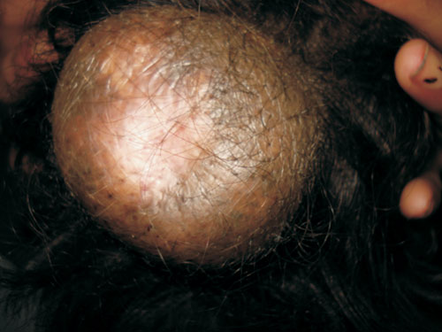
A 4-year-old female was admitted for bronchopneumonia. The child was also
noted to have a scalp neoplasm, which was present since birth but had
gradually increased to the present size (8X5.5 cm), with thick and
wrinkled covering skin (Fig. 1). The lesion was located in
the occipital region, was non-pulsatile, with no increase in size on
crying and no transillumination, thus ruling out a Neural tube defect. The
child had been developing normally with no neurological complaints or
similar family history. The patient also had a few pigmented nevi on the
back and the extremities. Neuroimaging of the brain was normal and the
biopsy showed numerous melanocytes in an intradermal location with
occasional melanophages and no cellular atypia. Child was referred to the
surgeons for operative management.
 |
|
Fig. 1 Giant pigmented nevus of scalp.
|
Giant congenital pigmented nevus shares the
pathological characteristics of common nevi and may invade the skin of any
region of the body. Its clinical importance is that its presence on the
head and neck region is usually complicated with neurocutaneous melanosis
(NCM), a rare phakomatosis consisting of congenital abnormal pigmentation
of the skin and meninges, and neurological features like epilepsy,
developmental delay, or focal neurological deficits may be present. The
meningeal lesions may undergo malignant change, with a very poor
prognosis. In NCM, the congenital melanocytic nevi are either giant (>20
cm in greatest dimension) or multiple (>3). Diagnosis is based on clinical
features and pathological examination. Treatment is radical or staged
excision. It must be differentiated from cutis verticis gyrate, in which
the skin lesions are symmetric, and complicated with proliferation of the
skin, hyperplasia of the periosteum and bone matrix.

