|
|
Case Reports Indian Pediatrics 2002; 39:588-592 |
||
|
Biliary Peritonitis: A Rare Presentation of Perforated Choledochal Cyst |
||
|
Mayur Maheshwari
Choledochal cyst is a relatively rare surgical entity. It is more common in Japan from where most of the cases were reported(1). Ruptured choledochal cyst is a rare clinical entity with the first case described by Weber in 1934(2). The incidence of perforation is around 2%(3,4). Here we are describing a case of ruptured choledochal cyst that presented as biliary peritonitis. Case Report A 6-year-old girl presented with fever and jaundice of 15 days duration, and vomiting and abdominal distention of 3 days duration. The patient was in a poor general condition with severe tachycardia, hypotension, pallor, respiratory distress, deep jaundice, dehydration, huge abdominal distention, and free fluid in abdomen. Paracentesis showed frank bile. The child was resuscitated, Ascitic tap was done (2 liters over 48 h) which relieved the distension and respiratory distress. Investigations revealed a serum bilirubin of 6.5 mg/dl (direct 5.0 mg/dL), alkaline phosphatase of 13.8 KA units; SGOT of 98 units; and PT of 20 sec (control of 24 sec). Ultrasonography (USG) abdomen showed moderate enlargement of liver with minimal intrahepatic biliary duct dilatation, fusiform dilatation of the CBD measuring 19 mm in maximal diameter, normal gall bladder and massive free fluid. Ascitic fluid showed bile salts and bile pigments. After stabilization, the patient was taken up for exploration, which showed biliary fluid of about one liter, a perforation on the anterior wall of the choledochal cyst of about 1 cm in size. This cyst was looking markedly inflamed and edematous. Liver was grossly normal. Simple T-tube drainage of the common bile duct (CBD) was done without excision of the cyst. Simultaneously, a feeding jejunostomy was done for feeding bile. Post-operatively, the patient drained about 400-500 cc of bile over 24 hours daily, which was fed to the patient through the jejunostomy. A T-tube cholangiogram, done on the 10th post-operative day, showed a dilated CBD, dilated CHD and cystic duct, with passage of the dye into the duodenum (Fig. 1). Intra-hepatic biliary radicles were normal and junction of CBD with the pancreatic duct appeared longer. T-tube was intermittently clamped, and bilirubin levels remained low. The T-tube was removed on the 14th post operative day. Jejunostomy was also removed. The wound healed and the patient was discharged. At 6 months follow-up, the patient was asymptomatic. USG showed a cyst of size 15 mm with no dilatation of the intrahepatic radicles. The patient was still awaiting definitive surgery and was lost to follow-up after 6 months. Discussion Cyst rupture with bile peritonitis is a rare complication of choledochal cyst. The reported incidence of perforated choledochal cyst varies from 2-18%(5-7). The exact cause of rupture is not definitely known. Distal obstruction is an important etiological factor along with primary weakness of duct wall(8). Trauma(9), association with pregnancy(10), anomalous pancreatico-biliary duct system(11), and continuous reflux of pancreatic juice in cyst weakening the wall(12) have been implicated in few cases. In infants commoner causes of bile peritonitis include spontaneous perforation of CBD, which usually occurs at the junction with cystic duct(13). Cystic duct perforation has also been reported(14). Rupture of the cyst is more common in infancy and rare in older children. Onset of symptoms is usually acute with vomiting, abdominal distension, features of toxemia, signs of shock, with or without icterus. The sub-acute form has been designated as "biliary ascites" with features of ascites, jaundice, fluid hernia and acholic stools. The acute form may raise the suspicion of rupture of a hollow viscus. In infants an additional differential diagnosis would be spontaneous perforation of some part of extrahepatic biliary apparatus such as CBD or cystic duct. Usually, the definitive diagnosis is possible at laparotomy. Ultrasonography can provide a diagnosis of ruptured cyst although it is difficult as cyst wall collapses after perforation. USG also provides information regarding the status of liver, presence of dilated intrahepatic radicals, presence of stones, diameter of pancreatic duct and presence of ascites. Definitive preoperative diagnosis can only be provided by hepatobiliary scintigraphy(15). Scintigraphy with delayed films can demonstrate leak from extrahepatic biliary ducts into general peritoneal cavity. Percutaneous transhepatic cholangiography (PTC) and Endoscopic retrograde cholangiopancreaticography (ERPC) may also be useful in diagnosis of ruptured cyst
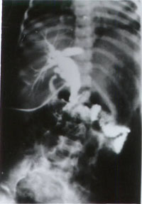
Fig. 1. T-tube cholangiogram showing dilated CBD, dilated CHD, cystic dilatation of cystic duct with passage of dye in duodenum. On exploration, the abdominal cavity was full of bile and pus flakes. There was a definite cyst of common bile duct (CBD) with perforation on the anterior wall. Grossly, the liver appeared to be normal. The wall of choledochal cyst was markedly inflamed and edematous. On exploration spontaneous perforation of CBD might give an appearance of perforated choledochal cyst. The picture was definitely not of localized bile collection so as to raise the suspicion of a pseudocyst. It is important to differentiate ruptured choledochal cyst from spontaneous perforation of extrahepatic biliary tree with walled off collection forming a pseudocyst which occurs almost exclusively in infants less than 20 weeks of age(13). Management of this condition requires simple drainage which usually leads to closure of perforation. Diagnosis of ruptured cyst may be difficult even at laparotomy because of collapse of the cyst wall, the usual location of the perforation is on the posterior wall. In view of the poor general condition and the local findings, it was decided not to do an extensive, time consuming dissection of cyst excision and biliary reconstruction in an emergency procedure. A life saving minimal procedure of external T-tube drainage was performed. Tube jejunostomy was done for postoperative bile feeding so that early enteral nutrition could be established to do complete excision and biliary-enteric anastomosis at the same time. A simpler procedure like T-tube drainage might be a better option for the time being to tide over the crisis and definitive surgery can be done at a later date(7). There are reports of ruptured choledochal cyst being managed primarily by cyst excision and biliary-enteric anasto-mosis(16). It is important to do a liver biopsy, as it is crucial for prognosis and follow up of the patients. It varies from being normal to mild inflammatory infiltrates of portal tracts and periportal fibrosis to frank cirrhosis. If expertise and facilities are available, endoscopic procedures such as ERCP, sphincterectomy and stenting or nasobiliary drainage can be performed as temporary measures to deal with the acute crisis. It is useful to feed the same bile to replenish the enterohepatic circulation and increase the bile acid pool. This requires a postpyloric nasogastric tube or a feeding tube distally(17). We did tube jejunostomy to feed bile drained by T-tube and could establish full enteral feeds early. Ruptured choledochal cyst is a rare pediatric surgical entity. This may be confused with rupture of a hollow viscus and can be missed intraoperatively, unless specifically looked for. In a infant it should be differentiated from pseudocyst due to localized bile collection in relation to ruptured extrahepatic biliary tree. This condition although rare should be kept in mind while dealing with any patient with bile peritonitis. Presence of any fluid suggestive of bile should prompt a surgeon to evaluate the biliary tree. In emergency situations such as cited above, external tube drainage is a simple, safe and life saving procedure. Excision of the cyst with biliary enteric anastomosis can be done at a later date on an elective basis with long term follow-up. Contributors: MM collected the data and drafted the paper; he will act as the guarantor for the manuscript. BRP and BKL both provided guidance and revised the draft. Funding: None. Competing interests: None stated.
| ||
| References | ||
|
![]()