A 3-month-old girl was brought to us with complaints of fever and
breathlessness for two days. On examination, child was afebrile, had
severe tachypnea, hypoxia and firm hepatosplenomegaly. There were
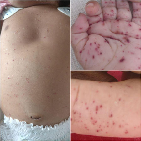
diffuse cutaneous lesions – hemorrhagic papules and vesicles on the
scalp, face, trunk and extremities, including palms and soles (Fig.
1). The mother reported the lesions to be present at birth but were
progressively increasing in number. The child had been evaluated for the
lesions in the newborn period and a presumptive diagnosis of Diffuse
neonatal hemangiomatosis was made due to the vascularized appearance of
the cutaneous lesions; a biopsy was not attempted. As the child was
otherwise asymptomatic, she was advised to follow-up.
 |
|
Fig. 1 Diffuse hemmorhagic papules
over trunk, palms and soles.
|
On laboratory evaluation, she had anemia and
hypoproteinemia. Computed tomography of chest revealed diffuse ground
glass appearance suggestive of Interstitial lung disease. Skin biopsy
demonstrated focal collection of cells with grooved nuclei.
Immuno-histochemistry revealed S100+ and CD1a+ cells. The final
diagnosis was congenital Langerhans Cell Histiocytosis (LCH), possibly
with progression from single system (skin) to multi-system (liver,
spleen, lung) involvement. The child was treated as per 2009 Histiocyte
Society guidelines for LCH protocol. On follow-up, the cutaneous lesions
completed regressed and child could be weaned-off oxygen.
Neonatal/congenital LCH is defined when it presents
within the first 4 weeks of life (irrespective of age at diagnosis). The
largest cohort study of neonatal LCH reported that 61 out of 1,069 LCH
patients (6%) met the criteria for neonatal LCH [1]. Skin lesions are
the most common initial manifestation, irrespective of disease extent at
diagnosis.
Differential diagnosis of hemmorhagic skin lesions
presenting in the neonatal period include cytomegalovirosis, candidiasis,
varicella, herpes simplex, neonatal toxic erythema, infantile
acropustulosis, pigmentary incontinence, eosinophilic pustular
folliculitis, neonatal erythropoiesis, disseminated neonatal
hemangiomatosis and congenital leukemia cutis. The presence of firm
hepatosplenomegaly and ILD (due to deposition of histiocytes) should
raise the index of suspicion towards LCH. Disseminated neonatal
hemangiomatosis can be a close mimic as they can present with visceral
involvement, and can be best differentiated by skin biopsy [2].
Isolated cutaneous LCH has a high tendency for
spontaneous regression, and has been described as Congenital
self-healing reticulohistiocytosis. However, approximately 60% can
progress to multisystem involvement, necessitating close follow-up.
References
1. Minkov M, Prosch H, Steiner M, Grois N, Pötschger
U, Kaatsch P, et al. Langerhans cell histiocytosis in neonates.
Pediatr Blood Cancer. 2005; 45:802-7.
2. Rubio González B, García Bracamonte B, Ortiz
Romero P, Postigo Llorente C, Vanaclocha Sebastián F. Multisystemic
langerhans cell histiocytosis mimicking diffuse neonatal hemangiomatosis.
Pediatr Dermatol. 2014;31;e87-9.


