|
|
|
Indian Pediatr 2018;55:
603-604 |
 |
Empyema Due to Thoracic Migrating
Appendicolith
|
|
Lakshmi Sundararajan 1,
K Prabhu1,
Venkateswari Ramesh2
and Janani Sankar2
From the Departments of 1Pediatric Surgery, and 2Paediatrics;
CHILDS Trust Medical Research Foundation, Kanchi Kamakoti CHILDS Trust
Hospital, Chennai, India.
Correspondence to: Dr Lakshmi Sundararajan, Department of Pediatric
Surgery, Kanchi Kamakoti CHILDS Trust Hospital, 12-A Nageswara Road,
Nungambakkam, Chennai 600 034, India.
Email: [email protected]
Received: January 19, 2017;
Initial review: May 18, 2017;
Accepted: March 28, 2018.
|
Background: Retained appendicolith following appendicectomy, and can
cause recurrent abscess in the abdomen and retroperitoneum. Case
characteristics: 11-yr-old boy who presented with subpulmonic
abscess and pneumonia following appendicectomy for perforated
appendicitis. Observations: Thoracotomy revealed a thick walled
subpulmonic abscess surrounding an appendicolith along with a rent in
the posterolateral aspect of the diaphragm. Message: In children
presenting with pus collections and a history of recent appendicectomy,
the possibility of a migrating appendicolith should be considered.
Keywords: Appendicitis, Appendicectomy, Complications.
|
|
D
ropped appendicolith can occur as a consequence
of non-retrieval of stone from peritoneal cavity during open or
laparoscopic appendicectomy. Such events are known to present with
delayed abscess in abdominal locations [1]. We report a child who
presented to us with subpulmonic abscess and pneumonia due to a dropped
appendicolith, following perforated appendicitis.
Case Report
An 11-year-old boy, previously hospitalized
elsewhere, had undergone emergency laparotomy and open appendicectomy
for perforated appendicitis with peritonitis. He had been treated with
broad spectrum antibiotics for ten days and discharged. During that
period he had some cough, but a normal chest X-ray. Two weeks
following surgery, he was brought to our hospital with intermittent high
grade fever for 3 days. On examination, he was febrile and tachypneic,
but not hypoxic. Air entry was decreased and there was stony dullness on
percussion in left hemithorax. Laboratory work-up revealed neutrophilic
leucocytosis, anemia (Hb 8.5 g/dL), and mild thrombocytosis (platelet
count 4.57 lakh/mm 3). He was
treated with broad spectrum antibiotics – Piperacillin-Tazobactam and
Vancomycin– in view of probable nosocomial infection. Chest X-ray
showed homogenous opacity in left middle and lower zones.
Ultrasonography (USG) of the chest showed thick multiloculated turbid
fluid collection in posterolateral aspect of left pleural cavity. Hence,
a diagnosis of complicated pneumonia with left empyema was considered.
USG abdomen done to rule out subdiaphragmatic abscess was normal.
Diagnostic thoracentesis resulted in a dry tap.
The child underwent a Video-assisted thoracoscopic
surgery (VATS), which showed loculated empyema, and thickened visceral
and parietal pleura with interpleural adhesions. Decortication was done
over upper lobe resulting in good lung expansion; however, we were
unable to clear the peel around lower lobe. Contrast enhanced computed
topography (CECT) of chest showed collapse of left lower lobe, mild to
moderate pleural fluid with air pockets and a radio-opaque shadow
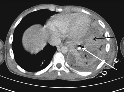
measuring (7x5 mm) in basal segment of left lower lobe Fig. 1.
Flexible bronchoscopy showed a normal trachea-bronchial tree with no
foreign body.
 |
|
Fig. 1 CECT chest showing collection
(black arrow) and a hyper-intense shadow suggestive of foreign
body (white arrow) in left hemithorax.
|
At thoracotomy, a thick walled subpulmonic abscess
surrounding an appendicolith (0.7×1.5 cm) was found. A rent in the
posterolateral aspect of diaphragm was noted (Web Fig. 1).
The rent in diaphragm was repaired and extensive decortication was done.
Appendicolith analysis was positive for calcium oxalate and carbonate.
Pus in the subpulmonic space and stone revealed growth of
Extended-spectrum Beta Lactamase Escherichia Coli, sensitive to
Piperacillin-Tazobactam. Post thoracotomy, fever settled and left lung
expansion improved. Child was discharged on oral co-trimoxazole for two
weeks after completion of two weeks of parenteral
piperacillin-tazobactam. At six months follow-up, he had full lung
expansion, and was doing well.
Discussion
An appendicolith is found in approximately 12-30% of
patients with appendicitis [2]. An appendicolith, also known as fecolith
or stercolith, is an inspissated fecal mass with calcium phosphate and
organic debris deposited around it. These are usually subcentimetric;
those larger than 2 cm are termed giant appendicoliths [3]. Formation of
abscess around such appendicolith occurs due to the bacteria within it
acting as a nidus of infection [4]. The time interval between
appendicectomy and the diagnosis of ectopic appendicolith may range from
ten days to few years [4].
Retained appendicoliths are most commonly found in
pelvis or Morrison’s pouch [5]. Pneumoperitoneum and positioning used
during laparoscopy can result in the appendicolith moving to unusual
sites. Abscesses have been reported in the retroperitoneum, perihepatic
region, and subhepatic, tubo-ovarian, psoas, pelvic and gluteal regions
[1,6]. A single case report of a migrating appendicolith with
diaphragmatic perforation resulting in empyema has been reported [7].
It is interesting that the appendicolith in our patient migrated
from the abdomen, perforating the diaphragm without causing any
significant subdiaphragmatic abscess.
X-rays can detect only 10-15% of appendicoliths.
USG and CT scan are diagnostic modalities of choice. In USG, calcified
appendicoliths are seen as hyperechoic foci. CT is more sensitive,
detecting even noncalcified fecoliths. On CT, appendicoliths appear as
areas of high attenuation, as laminated bodies with gas in centre or
homogenous opacity [8]. CT procedures to localize retained
appendicoliths preoperatively have been described [9]. In our patient,
appendicolith was initially not identified on X-ray and USG. CT
chest revealed a radiopaque shadow that was later confirmed as
appendicolith.
Retrieval of the appendicolith is important to
prevent future recurrent abscesses [1,10]. This can be done as open
surgery along with drainage of the abscess; percutaneous methods with
radiological guidance have also been described [1]. Prevention is by
meticulous attention during appendicectomy with retrieval of appendix as
well as any appendicoliths during surgery. Retrieval bags at laparoscopy
are useful as the lith may crumble during instrumentation [4].
We suggest that the possibility of a missed migrating
appendicolith should be considered in children who present with empyema
following appendicectomy.
Acknowledgements: Dr Nivedhana, Department of
Microbiology, Kanchi Kamakoti CHILDS Trust Hospital.
Contributors: All authors were involved in case
management and drafting the manuscript, and approved the final version
of manuscript.
Funding: None; Competing interest: None
stated.
References
1. Schraffl D, Frima H, Villiger P. A four-year
hospital journey for a lost and migrating appendicolith.Case Rep Surg.
2015; doi:10.1155/2015/832434.
2. Singh M, Kadian YS, Rattan KN, Jangra B.
Complicated appendicitis: Analysis of risk factors in children. Afr J
Paediatr Surg. 2014;11:109-13.
3. Garg PK, Jain BK, Rathi V, Mohanty D, Vaibhaw K.
Giant appendicolith. Indian J Gastroenterol. 2011;30:243.
4. Ajitha MB, Yethadka R, Sharath Kumar KL. Dropped
appendicolith: Complications and management. Int J Biomed Res.
2015;6:65-70.
5. Kim N, Reed WP, Abbas MA, Katz DS. CT
Identification of Abscesses after dropped appendicoliths During
Laparoscopic Appendectomy. Am J Roentgenol. 2004;182:1203-5.
6. Lambo A, Nchimi A, Khamis J, Khuc T.
Retroperitoneal abscess from dropped appendicolith complicating
laparoscopic appendectomy. Eur J Pediatr Surg. 2007;17:139-41.
7. Betancourt SL, Palacio D, Bisset GS. The
‘wandering appendicolith’. Pediatr Radiol. 2015~45:10914.
8. Aiken JJ, Oldham KT. Acute Appendicitis. In:
Kliegman RM, Stanton BF, St. Gema JW, Schor NF, Behrman RE, editors.
Nelson Textbook of Pediatrics, Philadelphia: WB Saunders Co, 20th ed.
2011. p.1889-93.
9. Lossef SV. CT-guided Kopans hookwire placement for
preoperative localization of an appendicolith. Am J Roentgenol.
2005;185:81-3.
10. Buckley O, Geoghegan T, Ridgeway P, Colhoun E,
Snow A, Torreggiani WC. The usefulness of CT guided drainage of
abscesses caused by retained appendicoliths. Eur J Radiol. 2006;60:80-3.
|
|
|
 |
|
