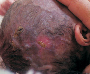
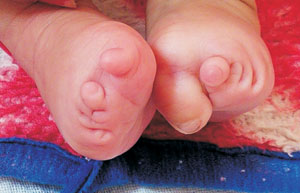
A 2-month-old girl, born to a non-consanguinously married
parents, presented to us with presence of an open wound at
birth which healed with scarring and hair loss, along with
abnormalities involving toes of both feet. The lesion was an
atrophic scar tissue with a rough and heterogeneous
appearance. Physical examination revealed localized alopecia
with dimensions of 1.8 × 4 cm (Fig. 1a), along
with prominent scalp veins. Cutis marmorata was present all
over body. Oligodactyly and digital nubbins were present in
bilateral feet (Fig. 1b).
 |
 |
|
Fig.1 (a) Alopecia cutis
congenita of scalp, prominent scalp veins;
(b) Oligodactyly and presence of digital nubbins.
|
Based on the classical clinical
presentation of aplasia cutis congenita of scalp, cutis
marmorata, prominent scalp veins, and limb abnormality, a
diagnosis of Adams Oliver syndrome was considered.
Histopathology of hairless atrophic patch showed loss of
rete ridges, collagen deposition and loss of skin
appendages. Hemogram, liver function tests, kidney function
tests, serum electrolytes and Chest X-ray were
normal.
Adams Oliver Syndrome is a rare and
clinically heterogeneous anomaly characterized by the
combined occurrence of congenital scalp defects and terminal
transverse limb defects. It includes aplasia cutis congenita,
variable limb defects, and associated anomalies ranging from
skin tags to lymphedema. Other system anomalies and
malformations such as cardio-vascular, respiratory and
orofacial defects have also been reported. With conservative
therapy to prevent secondary infection and consequent tissue
damage, most small defects of scalp heal well during the
first few months of life. Larger and obvious scars can be
treated with plastic surgical reconstruction. The lesions of
cutis marmorata may fade with time during first year of life
due to skin thickening and maturation.


