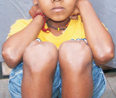
A nine-year-old boy presented with multiple painful nodular lesions on the
extensor aspect of bilateral elbow and knee joints since 15 days (Fig.
1). There was no sore throat, pyoderma, arthralgia, abdominal
pain, drug intake or Koch’s contact. On enquiry, fever and exertional
dyspnea were present since 5 days. Grade III/VI pansystolic murmur was
present in the mitral area. Chest x-ray and ultrasonography of abdomen
were normal. Mantoux test was negative. Antistreptolysin titre,
erythrocyte sedimentation rate and C-reactive protein were elevated.
Echocardiography showed moderate mitral regurgitation. Histopathology of
the nodular lesions was consistent with erythema nodosum (EN). With a
diagnosis of rheumatic heart disease and active carditis, benzathine
penicillin prophylaxis and aspirin were started. On follow up after 3
weeks, the nodules had disappeared.
 |
|
Fig. 1 Nodular lesions on the extensor
aspect of bilateral elbow and knee joints. |
EN is a symmetric inflammatory process involving the
subcutaneous fat that causes tender, erythematous nodules. Sites
involved are pretibial (most common), extensor surfaces of forearm, legs,
thighs, and trunk. The lesions do not ulcerate and resolve
without atrophy or scarring in one to two months. EN is a cutaneous
immune-mediated (type IV delayed hypersensitivity) reaction
to a variety of antigens. Commonly associated conditions include
streptococcal infection, tuberculosis, sarcoidosis, sulphonamides,
amoxicillin, inflammatory bowel disease, lymphoma, amoebiasis, giardiasis
and viral infections (hepatitis B & C, herpes simplex, HIV and EBV).
Common differential diagnoses include infectious panniculitis, lupus
panniculitis, cold panniculitis, leukemic infiltrates, necrobiosis
lipoidica, lipodystrophies and scleroderma. Management includes treatment
of underlying disorders and supportive care i.e. bed rest, avoiding
contact irritation of affected areas, non-steroidal anti-inflammatory
drugs for pain and systemic steroids.


