1.gif (1892 bytes)                     

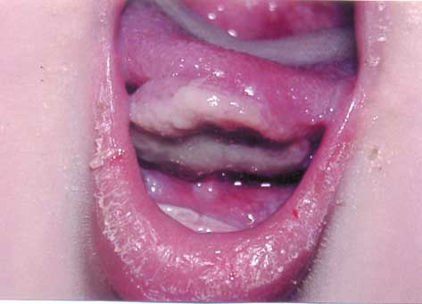
Images

Indian Pediatrics 2008; 45:609

Ulcerative Traumatic Granuloma of the Oral Cavity


An 8 month-old infant boy with cerebral palsy presented with a large exophytic lesion under the tongue with erythema surrounding a removable, fibrinopurulent membrane and granulation tissue proliferation present for several weeks (Fig. 1). He was unable to suckle for feeding. Laboratory tests were normal. Biopsy was suggestive of distinctive ulcerative traumatic granuloma with stromal eosinophilia (Riga-Fede disease). Contact of the tongue with the sharp, newly erupted mandibular teeth was apparent. Grinding the teeth edges and covering with resin resulted in rapid resolution within 5-10 days.

Fig. 1. Ulcerative traumatic granuloma on the anterior ventral part of the tongue; note sloughing necrotic pseudomembrane, and dry lips.

Riga-Fede disease, a variation of the traumatic granuloma, may occur after acute injuries with sharp foodstuffs, biting, or mastication. This unique type of chronic granulomatous ulceration with stromal eosinophilia is a deep, pseudoinvasive, inflammatory reaction seen in infants following chronic trauma from neonatal or primary teeth. Differential diagnosis may include pyogenic granuloma, ulcerative carcinoma, and lymphoma. Biopsy provides definitive diagnosis. Removing the source of trauma is sufficient. Failure to diagnose and treat RFD results in dehydration and inadequate intake for the infant.

Taghi Azizi,
Mohammad Hosein Kalantar Motamedi,
Departments of Pathology and
Trauma Research,
Baqiyatallah University of Medical Sciences,
Tehran, Iran.
.

Home

Past Issue

About IP

About IAP

Feedback

Links

 Author Info.

  Subscription