A 10-year-old male presented with inability to walk alone due to
weakness of both upper and lower limbs for one year along with slurred
speech and drooling of saliva for three years. There was no history of
convulsion, jaundice, measles or recent vaccination. He was emotionally
labile and had slurred speech. There was hypertonicity of all four limbs
with dystonia, bilateral extensor plantar reflex, ataxia and intention
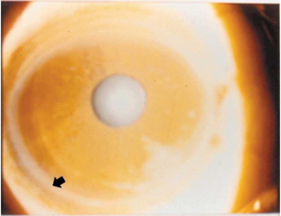
tremor. Liver was just palpable. Examination of eyes revealed presence
of Kayser-Fleischer (KF) ring (Fig. 1) bilaterally. On slit lamp
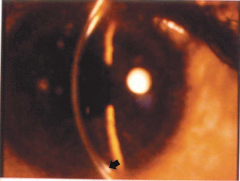
examination, the ring (3 mm in width) appeared as a golden-brown deposit
at the level of the Descemetís membrane of cornea (Fig. 2). A
diagnosis of Wilsonís disease was made. The serum ceruloplasmin was 3.5
mg/dL, 24 hours urinary copper was 120 Ķg/dL. The patient was treated
with d-penicillamine and the KF ring disappeared.
 |
 |
Fig. 1. Kayser-Fleischer ring in cornea.
|
Fig. 2. Slit lamp examination reveals golden brown deposit in
Descemetís membrane. |
KF ring is the pathognomic sign of Wilsonís disease.
It may be absent in young patients with hepatic symptoms but is always
present in patients with neurological symp-toms. It is present in 98% of
neurologically symptomatic and 40% of asymptomatic patients with
Wilsonís disease. The ring appears as a golden-brown to green deposit at
the level of the Descemetís membrane of cornea. It ranges from 1 to 3 mm
in width, progressing centrally. It is usually bilateral. Deposition
begins in the superior cornea, than the inferior cornea, and eventually
forms a complete ring. Treatment with d-penicilla-mine causes the ring
to disappear, in the reverse order of its formation.
Sumana Datta,
Himadri Datta,
Department of Pediatrics,
Calcutta National Medical College and
Regional Institute of Ophthalmology,
Kolkata,
India.
E-mail:
[email protected]
