|
|
Images in Clinical Practice Indian Pediatrics 2004;742 |
||||
|
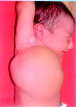
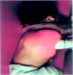
Lymphomatous Malformation of the Chest Wall |
||||
|
Previously known as cystic hygroma, these lesions are presently classified as benign congenital lymphomatous malformations consisting of ectatic channels filled with lymphatic fluid. They are usually located in the neck, axilla or mediastinum and they would never regress spontaneously. A Doppler ultrasound should be done to detect any vascular component of the lymphangioma and a contrast CT to delineate the depth of the lesion. The lesions can increase in size during any systemic infection or due to intralesional bleeding. A complete surgical resection is the treatment of choice. Subtotal resection either due to its presence at strategic site (involving the blood vessels/nerves as in the neck), or due to its deeper extent, may lead to recurrence. Local injection of sclerosants viz 100% ethanol or OK-432 ( a killed strain of group A Streptococcus) may be beneficial in a macrocystic lymphomatous malformation. However, this may leave scarring in the skin. Arti Maria, |
![]()