|
CHOLEDOCHAL CYSTS IN INFANTS AND CHILDREN |
Ujjal Poddar, B.R. Thapa, Mohinish Chhabra, K.L.N. Rao*, S.K. Mitra*, J.B. Dilawari, and Kartar Singh
From the Division of Pediatric Gastroenterology, Department of Gastroenterology and Department of Pediatric Surgery*, Postgraduate Institute of Medical Education and Research, Chandigarh 160012.
Reprint requests: Dr. B.R. Thapa, Additional Professor, Division of Pediatric Gastroenterology, Department of Gastroenterology, Post Graduate Institute of Medical Education and Research, Chandigarh 160012.
Manuscript received: January 30, 1997; Initial review completed: March 4, 1997;
Revision accepted: January 1, 1998
Abstract:
Objective: To study the clinical spectrum and managment of choledochal cyst
in children below
12
years of age. Design: Descriptive study. Setting: Tertiary care hospital. Methods: Twenty
three children with choledochal cysts were managed between January
1991 to September 1997 and their clinical details, investigations and management were recorded. Choledochal cyst was diagnosed by ultrasonography and confirmed by ERCP or
peroperative cholangioram (POC).
Children were treated with antibiotics and/or percutaneous transhepatic biliary drainage
if
there
was cholangitis and subsequently subjected to surgery (excision of the cyst and jejunal loop interposition hepaticoduodenostomy). Results: The median age of these children was
3 years with an almost equal sex ratio. Predomillant presentation was jaundice in 18, pain abdomen in 15, fever in 12, and lump abdomell in 9
cases. The classical triad of jaundice, pain and lump was
present in only 4 cases.
ERCP
conducted in
7 and POC in 14 cases yielded positive findings in all. Clinically there were two distinct forms of presentation: (i) infantile
form (≤
1
year) comprised 9 infants which presented with jaundice in all, acholic stool in 6, lump abdomen in 4 but only one had classical triad; and (ii) childhood
form (> 1 year) presented with pain abdomen in 12 and jaundice and cholangitis in 9
subjects each. Type
I
cyst was seen in
20 and type IVa
in 3. Two children refused surgery, and the rest underwent surgery. Three infants died after
surgery, the remaining 18 were alive and well on follow-up (median
25
months). Secondary
biliary cirrhosis was seen in
6, extra hepatic biliary artresia in 2 and congenital hepatic fibrosis in 1 on histology. Conclusions: Choledochal cysts present in two clinically distinct forms. Infantile form is an important cause of c1lOlestasis of infancy. Early diagnosis and referral is essential to prevent complications and death, and prognosis after surgery is good.
Key words: Choledochal cyst; Endoscopic retrograde; cholangio-pancreatography.
CHOLEDOCHAL cyst, a localized
aneurysmal dilatation of the extrahepatic and/or intrahepatic biliary tree, is an uncommon cause of obstructive jaundice and is diagnosed mainly in the pediatric age
group(1). Vater in 1723 had given the first published description of choledochal cyst but in 1852 Douglas provided the first clinical description of a patient with choledochal cyst. Since then till 1990 only
3000 cases
have been reported(2). More
than two third of these reports are from Japan, and only two reported clinical series of choledochal cysts in children are from India(3,4). One report is a decade old(3) and another one(4) has compared children
of > 6 years with adults, from which we do not get the true clinical spectrum of choledochal cysts in children. We, there- fore, studied the clinical spectrum and management of choledochal cysts in Indian children (≤ 12 years).
Subjects and Methods
Twenty three cases of choledochal cysts were managed at our center from January 1991 to September 1997. Their clinical details, investigations and management were recorded. Ultrasound examination of abdomen was done in all of them. Presence of a dilated common bile duct, with or without dilated intrahepatic ducts was labelled as choledochal cyst and the diagnosis was confirmed by peroperative cholangiogram (POC) or endoscopic retrograde cholangiopancreatogram (ERCP). Esophagogastroduodenoscopy was done when there was a suspicion of portal hy- pertension either clinically (splenomegaly) or sonologically and repeated on follow up if there were esophageal varices. Children with cholangitis were treated with antibiotics and percutaneous transhepatic biliary, drainage (PTBD) was done if there was no
response to antibiotics. After controlling cholangitis, all these children were offered surgery. The surgical procedure included excision of the cysts and jejunal loop interposition hepaticoduoclenostomy.
In
cases of extrahepatic biliary atresia (EHBA)
with choledochal cyst, portoenterostomy was done. Wedge liver biopsy and sect- ed specimens were subjected to 'stologic examination. Cysts were classifed accord- ing to Todani et al.'s classification(S): Type I, solitary extrahepatic cyst; Type II, extrahepatic supraduodenal diverticulum; Type III, choledococele; Type IVa, extrahepatic and intrahepatic cysts;
Type
IVb
multiple extrahepatic cysts; and
Type
V,
Caroli's disease (multiple intrahepatic cysts).
Results
Twenty three children with choledochal cysts were diagnosed from 3093 admissions in our Pediatric Gastroenterology Center during the study period; comprising 0.7% of total admissions. Their median age was 3 years (range, 1 month to 12 years). Male and female ratio was 1.3:1. The duration of symptoms ranged from 6 days to 7 years (median 6 months)
and onset of symptoms ranged from 3 days of age to 9 years.
Their clinical features are summarized in Table I. Predominant presentations were jaundice, abdominal pain; and cholangitis. The classical triad of pain, jaundice and lump abdomen was present in only 17.4% of cases.
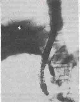
Investigations showed serum bilirubin and alkaline phosphatase were elevated in 18 children, transaminases in 7 and amylase in 2. Ultrasound could pick up choledochal cysts in all cases. ERCP could be done in 7 (youngest 18 months old) cases and in 6 it was suggestive of
Type
I
cysts and in one child
Type
IV a
cysts (Fig. 1). Secondary biliary cirrhosis was
documented in 6 children on preoperative wedge biopsy, two of
them died. In one infant repeat percutaneous biopsy 8 months
after surgery had shown regression of biopsy changes, other 3
were asymptomatic
on follow up but continued to have firm hepatomegaly. Esophageal varices were detected in 4; in one child they disappeared soon after surgery (due to decompression of portal vein caused earlier by cyst), but in other 3 they were persisting on follow up (1 due to congenital hepatic fibrosis, 2 due to secondary biliary cirrhosis).
TABLE I
Clinical
Features and Comparison Between Infantile and Childhood Form
|
Feature
|
Overall |
Infantile |
Childhood |
|
Median age (mo) |
36 |
5.5 |
63 |
|
Median duration of symptoms (mo) |
6 |
2 |
20 |
|
Jaundice |
18 |
9 |
9 |
|
Pain abodmen/irritability |
15 |
3 |
12 |
|
Fever |
12 |
4 |
8 |
|
Lump abdomen |
9 |
4 |
5 |
|
Classical triad |
4 |
1 |
3 |
|
Cholangitis |
13 |
4 |
9 |
|
Acholic stool |
7 |
6 |
1 |
|
Secondary biliary cirrhosis |
6 |
4 |
2 |
|
Stone in the cyst |
4 |
0 |
4 |
|
Mean (SD) serum bilirubin (mg/dl) |
6.3 (5.9) |
10.8 (6.5) |
3.5 (3) |
 |
|
Fig. 1. ERCP showing both intrahepatic (arrow) and extrahepatic
cysts (type IVa) in a 12 year old girl. |
Clinically there were two distinct forms of presentation in our children: infantile (≤1 year) and childhood or so called adult from (> 1 year). Significant differences
in presentation
(Table
I) were cholestatic jaundice
with acholic stool in infants (p < 0.01) and pain abdomen in children
(p < 0.01). Seven (50%) children presented with abdominal pain
associated with jaundice. One infant presented with perforated choledochal cyst who died after surgery due to septicemia. Two of our infants had associated EHBA diagnosed on the basis of liver biopsy, POC and histology of resected specimens. Both these cases had onset of symptoms within 1 week of life. Portoenterostomy
was done in both and they remained asymptomatic on follow-up. One
child had associated congenital hepatic fibrosis (CHF). Mean serum bilirubin was significantly higher in infantile form (p < 0.01), but alkaline phosphatase, SGOT and SGPT were not discriminatory. Death occurred only in child with the infantile form.
Anatomically, type I choledochal cyst was seen in 20 (87%) children and
Type
IVa
in 3 (13%). We did not encounter
Types
II,
III and V
in any of our children. Out of 13 children with cholangitis, 2 required PTBD and other 11 improved with antibiotics. Two children refused surgery, the otJ1er 21 cases underwent surgery; one of them was subjected to internal drainage in a peripheral hospital. This case presented to us 4 years later with variceal bleed due to CHF. We have operated 20 children, two infants died in the immediate post operative period due to anastomotic leak and septicemia, respectively. Another infant died of cholangitis 2 months after surgery. The other 18 cases were alive and well on follow up (median 25 months, range 1 to 80 months).
Discussion
Choledochal cyst is a rare congenital anomaly, more common in females, with an estimated incidence in Western populations of one in 100-1,50,000 live birth(6). The
incidence in our country is not known. In our center it is 0.7% of total admission which is 7 times more than adults, reported from another center of our country(2). The
lack of female preponderance observed by us has also been reported previously from our country(3), and in. some other Asiatic races(7).
We have seen two clinically distinct groups of patients; although this kind of presentation has been reported(8), if not stressed in the literature. Almost one third (39%) of our children were in the infantile group which has been reported in 18-29% of cases(1,9). Choledochal cyst should be seriously considered in infants with cholestatic jaundice as it is an important and treatable cause of cholestasis of in- fancy (9 out of 135 cases, i.e., 6% in our center). Ultrasound examination should be done promptly in all such infants to detect choledochal cyst. A delay in diagnosis may lead to fatal complications like spontaneous perforation, cholangitis and secondary biliary cirrhosis(10).
In our series also, delayed referral (median 2 months) was responsible for a high incidence of secondary biliary cirrhosis and subsequent mortality in infants. Similarly, choledochal
cyst should be suspected strongly in children presenting with pain abdomen especially when
combined with jaundice. Stringer
et al.(10)
in their series of 78 cases of choledochal cysts have shown that 50% of their children had jaundice associated with pain abdomen. We have also seen this combination in 50% of children. A delay in diagnosis in these children may lead to complications like cholangitis, cholelithiasis, secondary biliary cirrhosis and malignancy, which are reported mainly in adults(4).
Portal hypertension in patients with
choledochal cysts may occur either because of secondary biliary cirrhosis(9,11)
or direct compression by the cyst(12). One of our children had portal hypertension due t compression and another due to CHF
which is a known association of choledochal cyst(13,14). The prognosis
of
secondary biliary cirrhosis due to choledochal cyst is better than other causes. Regression of cirrhosis has been documented following cyst drainage(15) which we have also seen in one of our infant 8 months after surgery.
In 1959, preoperative diagnosis of choledochal cyst was possible in only 30% of
cases(1) but now it is possible in > 90% cases with the help of ultrasound and ERCP(9,16). Ultrasound is the initial screening investigation of choice and has been found to be very sensitive(9,17). However, the gold standard for morphological diagnosis and classification is still cholangiogram (endoscopic, percutaneous transhepatic or peroperative)(2,18). There are only few reports of use of ERCP in children with choledochal cyst(19,20). The youngest child with choledochal cyst in whom ERCP was done, was 2 years oId(21). We could do ERCP in 7 children and our youngest patient was 18 months old. We did not encounter any complication (except mild pancreatitis in one) and found it to be very useful in delineating the anatomy before surgery, there by obviating the need for POC. Another advantage of ERCP is placement of nasobiliary drain in presence of cholangitis which is safer than PTBD(22).
Treatment of choledochal cyst is surgical. Excision of the cyst is the treatment of choice, followed by establishment of bilioenteric communication(23). Commonly, the biliary reconstruction following excision has been carried out by Roux-en-Y hepaticojejunostomy. Hepatico-Roux-en-Y jejunostomy involves a high risk of peptic ulcer and fat malabsorption(24,25).. These drawbacks can be, overcome by jejunal interposition hepaticoduodenostomy. We have reported this procedure earlier(26) and it appears to be the most physiological method. With this technique only 3 of our
cases had complications (anastomotic leak and cholangitis).
In conclusion, choledochal cyst is not rare in Indian children. They present in two clinically distinct forms. The infantile form is an important cause of cholestasis of infancy. Early diagnosis and referral is essential, especially in infantile form to prevent complications and death. Prognosis is good after surgery.
1.
Alonso-Lej F, Rever WB, Pessagno DJ. Congenital choledochal cyst with a report of 2 and analysis of 94 cases. Abstr Surg 1959; 108: 1-30.
2.
Anand AC, Sahni P, Vashisht S, Tandon RK. Congenital biliary cysts in Indian adults. Am
J
Gastroenterol 1991; 86: 850- 853.
3.
Upadhyaya P, Upadhyaya P. Choledochal cyst. Indian Pediatr 1985; 22: 613-618.
4. Chaudhary A, Dhar P, Sachdev A, Kumar N, Vij JC, Sarin SK, et al. Choledochal cysts - differences in children and adults. Br
J
Surg 1996; 83: 186-188.
5.
Todani T, Watanabe Y, Narusue M, Tabuchi K, Okajima K. Congenital bile duct cysts: Classification, operative procedures, and review of thirty seven cases including cancer arising from choledochal
cyst. Am
J
Surg 1977; 134: 263-269.
6.
Howard ER. Choledochal cysts. In: Surgery of Liver Disease in Children. Ed. Howard ER. Oxford, Butterworth-Heineman, 1991; pp 78-90.
7.
Crittenden SL. McKinley MJ. Choledochal cyst: Clinical features and classification. Am
J
Gastroenterol1985; 80: 643-647.
8.
O'Neill JA, Templeton JM, Schnaufer L, Bishop HC, Ziegler MM, Ross AJ. Recent experience with choldeochal cyst. Ann Surg 1987; 205: 533-540.
9.Joseph VT. Surgical techniques and long
~
results in the treatment
of
choledochal cyst.
J
Pediatr Surg 1990; 25: 782-787.
10.
Stringer MD, Dhawan A, Davenport M, Mieli-Vergani G, Mowat AP, Howard ER. Choledochal cysts: Lessons from a 20 year experience. Arch Dis Child 1995; 73: 528- 531.
11.
Orenstein SR, Whitington PF. Choledochal cyst resulting in congenital cirrhosis. AmJ Dis Child 1982; 136: 1025-1026.
12.
Martin L W, Rowe GA. Portal hypertension secondary to choledochal cyst. Ann Surg 1979; 190: 638-639.
13.
Evans-Jones G, Cudmore R. Choledochal cyst and congenital hepatic fibrosis.
J
Pediatr Surg 1990; 12: 1259-1260.
14.
Lake DNW, Smith PM, Wheeler MH. Congenital hepatic fibrosis and choledochal cyst. Br Med
J
1977; 2: 1259- 1260.
15.
Yeong ML, Nicholson GI, Lee SP. Regression of biliary cirrhosis following choledochal cyst drainage. Gastroenterology 1982; 82: 332-335.
16.
Lopez RR, Pinson CW, Campbell JR, Harrison M, Katon RM. Variation in management based on type of choledochal cyst. AmJ Surg 1991; 161: 612-615.
17.
Hadidi A. Types I and III choledochal cyst. Preoperative diagnosis by ultrasound. AmJ Dis Child 1983; 137: 663c66
18.
Tan KC, Howard ER. Choledochal cyst: A
fourteen-year surgical experience with 36
patients. Br
J
Surg 1988; 75: 892-895.
19.
Weidmeyer DA, Stewart ET, Dodds WJ, Geenen JE, Vennes JA, Taylor AJ. Choledochal cyst, findings on cholangiopancreatography with emphasis on ectasia of the common channel. AJR 1989; 153: 969-972.
20.
Sharma AK, Wakhlu A, Sharma 55. The role of endoscopic retrograde cholangirpancreatography in the management of choledochal cysts in children.
J
Pediatr Surg 1995; 30: 65-67.
21.
Yamaguchi M. Congenital .choledochal cyst. Analysis of 1,433 patients in -the Japanese literature. Am
J
Surg 1980; 140: 653-657.
22.
Stanley J, Gobien RP, Cunningham J,
,
Andriole
J.
Biliary decompression: A institutional comparison of percutaneous and endoscopic methods. Radiology 1986;
158: 195-197.
23. Numez-Hoyo M, Lees CD, Herman RE. Bile duct cysts. Am
J
Surg 1982; 144: 295- 299.
24.
McArthur MS, Longmire WP. Peptic ulcer disease after choledochojejunostomy. Am
J
Surg 1971; 122: 155-158.
25.
Jensen SL, Nielsen OV, Lenz K, Nielsen ML. Fat malabsorption in patients with Roux-en-Y hapticojejunostomy. Surg Gynecol Obstet 1978; 147: 561-564.
26.
Narasirriha Rao KL, Mitra SK, Kochhar R, Thapa BR, Nagi B, Katariya 5, et al. Jejunal interposition hepatieoduodenostomy
for choledochal cyst. Am
J
Gastroenterol
1987; 82: 1042-1045.
|
|
| |
