Lead is an abundantly distributed heavy metal in our
environment which in higher concentrations is hazardous to the body [1].
Nervous system remains the most severely affected, effect being more
pronounced on growing children [2]. Common sources are lead based paint,
lead contaminated air, soil, dust, drinking water through lead soldered
pipes, lead coated vessels used for cooking, traditional medications and
certain cosmetics [1]. Absorbtion of lead varies depending on the
chemical form and the mode of exposure (ingestion > inhalation >transdermal).
The half life of lead in blood and soft tissues is 35 days as compared
to bones being 5-20 years. Bone stores release lead to the blood which
may add up to a toxicologically significant amount [3]. We report a boy
with lead encephalopathy, who required repeated chelation therapy.
A 7-year-old boy, presented with refractory status
epilepticus. He was the first born child of a non-consanguineous
marriage from Nilgiris, Tamilnadu, with unremarkable neonatal period. He
was developmentally and neurologically normal with no behavioural
problems and immunized upto date as per national immunization schedule.
There was no family history of seizures. The child was on traditional
medicines for about a year for vitiligo over lips and face. He developed
unilateral headache 10 days prior to the onset of seizures along with
intermittent abdominal pain and vomiting for which symptomatic treatment
was given. One such episode of vomiting was followed by right sided
focal seizures. The child was given parental anti-convulsants. However,
due to worsening of sensorium and uncontrolled seizures, he was put on
mechanical ventilation and started on multiple anticonvulsants. Child
was gradually stabilized and extubated.
Laboratory analysis showed leucocytosis with
polymorphic preponderance and microcytic hypochromic anemia. Serum Iron
was low - 21 mcg/dL [Normal 50-120 mcg/dL] though serum ferritin and
total iron binding capacity were within normal range. Liver functions,
renal functions and coagulation profile were normal. Cerebrospinal fluid
analysis showed mild leucocytosis, with minimally elevated proteins.
Cerebrospinal fluid Gene X pert for tuberculosis was negative.
Neuro-imaging was normal. Heavy metal screening of blood showed high
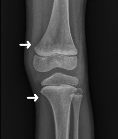
lead levels of 80.31 mcg/dL (acceptable upto 5 mcg/dL). Skeletal survey
showed lead lines over lower end of femur (Fig. 1). Parents were
screened and their blood lead levels (BLL) were within normal limits.
 |
|
Fig. 1 X-ray left knee of the
index patient showing lead lines (arrows) over lower end of
femur and upper end of tibia.
|
He underwent lead chelation therapy with
Dimercapto-succinic acid 30 mg/kg/day for 5 days followed by 20
mg/kg/day for 14 days. Other effective agents including Dimercaprol and
Edetate disodium calcium (CaNa2EDTA) could not be procured at that time.
Supplementation with Iron, vitamin D, zinc, vitamin C was done. He was
stabilized, anticonvulsants were gradually weaned off. BLL dropped to
38.08 mcg/dL. On review after 2 months, BLL showed a rise to 56.38 mcg/dL.
Child, however remained asymptomatic. Repeat chelation therapy was given
and BLL dropped further to 32.9 mcg/dL only to rise to 62.9 mcg/dL in 2
months. He has undergone four doses of periodic chelation at the time of
writing this report. He has been stable except for a few bouts of anger
outbursts for which he is on follow up with child psychiatrist. He is on
close follow-up and may require further chelation.
Lead is not known to serve any significant biological
function and deposition does not spare any organ in the body [1]. It has
high affinity for the skeleton and chronic exposure often sequesters
large proportion in the bones followed by the kidneys [4]. After a
period of initial exposure lead is redistributed to the soft tissues. If
cessation of exposure occurs at this juncture, there is a decrease in
the blood lead levels post the initial rise [5]. Bone, being a dynamic
tissue, undergoes remodelling throughout life which is regulated by a
wide range of hormones and local availability factors. Prolonged
exposure also results in slow release of lead from the bone stores over
a protracted period of time [4]. Children are at high risk of lead
poisoning as they are in a state of constant growth and development.
Moreover, the growing bones in children undergo perpetual remodelling
which allows lead to be regularly re-introduced into the blood stream
[6]. Chelation therapy brings down the blood lead levels acutely only to
rebound within weeks to months after treatment. Often, repeated courses
of chelation are required [5].
This case report emphasizes the need for long term
follow-up with periodic monitoring of lead levels in children with
chronic lead poisoning to assess the need for repeated chelation
therapy. Blood lead concentration may rise to toxic levels even after
removal of exposure due to constant re-distribution in a growing child.
1. Wani AL, Ara A, Usmani JA. Lead toxicity: A
review. Interdiscip Toxicol. 2015;8:55-64.
2. Keosaian J, Venkatesh T, D’Amico S, Gardiner P,
Saper R. Blood lead levels of children using traditional Indian medicine
and cosmetics: A feasibility study. Glob Adv Health Med. 2019;8:1-6.
3. Krishnaswamy K, kumar BD. Lead toxicity. Indian
Pediatr. 1998;35:209-16.
4. Pounds JG, Long GJ, Rosen JF. Cellular and
molecular toxicity of lead in bone. Environ Health Perspect. 1991;91:
17-32.
5. Hu H, Pepper L, Goldman R. Effect of repeated
occupational exposure to lead, cessation of exposure and chelation on
levels of lead in bone. Am J Ind Med. 1991;20:723-35.
6. Barbosa Jr F, Tanus- Santos JE, Gerlach RF, Parsons PJ. A critical
review of biomarkers used for monitoring human exposure to lead:
Advantages, limitations, and future needs. Environ Health Perspect.
2005;113:1669-74.

