|
|
|
Indian Pediatr 2019;56: 79- 80 |
 |
Aorto-pulmonary Collateral Masquerading as Cavity
|
|
Sakshi Sachdeva
Senior Resident, Pediatric Cardiology, Department of
Cardiology, Cardio Neuro Center (CNC), AIIMS, New Delhi, India.
Email:
[email protected]
|
|
An 8-year-old child presented to Pediatric cardiology outpatient
services with complaints of bluish discoloration of nails and lips with
poor growth since early childhood. On evaluation, child had low weight-
and height-for-age with central cyanosis and grade-2 pan-digital
clubbing. Oxygen saturation was 68% at room air. Cardiovascular
examination revealed single second sound without any murmur. Chest
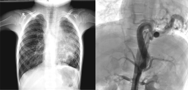
radiograph revealed pulmonary oligemia with left lower zone cavitary
lesion (Fig. 1a). There was no history of cough,
hemoptysis or tuberculosis contact, and evaluation of respiratory system
was normal.
 |
| (a) |
(b) |
|
Fig. 1 (a) Chest radiograph PA
view showing left lower zone cavitary lesion; (b) Angiographic
projection AP view showing a large tortuous aorto-pulmonary
collateral arising from celiac trunk and curving in left lower
zone masquerading as cavity on chest radiograph.
|
On echocardiography, the diagnosis confirmed was
tricuspid atresia and pulmonary atresia. Patient was planned for cardiac
catheterization to determine the source of pulmonary blood flow. Upon
catheterization, single large aorto-pulmonary collateral arising from
celiac trunk was noted, which was the sole source of pulmonary blood
flow (Fig. 1b). The tortuosity of collateral in
left lower zone of lung appeared like a cavity on chest radiograph.
Incidence of tuberculosis in congenital heart disease
is almost 2.5 times that of the normal population [1]. In children with
increased pulmonary blood flow, it is because of chronically wet lungs,
and in those with reduced pulmonary blood flow, it happens because of
ventilation-perfusion mismatch. Recognizing and treating tuberculosis is
important before cardiac surgery. In cyanotic congenital heart disease
with reduced pulmonary blood flow, radiological features mimicking
tuberculosis include apical caps and pseudo fibrotic lesions [2,3].
Collaterals give lacy reticular pattern with non-homogenous pulmonary
vascularity as collateral flow is non-uniform [4]. Aortopulmonary
collaterals in our patient radiologically suggested pulmonary
parenchymal pathology, sometimes they manifest with hemoptysis as well,
making the differentiation further difficult. Awareness of this possibly
may help the clinician to suspect and appropriately manage these
children.
Acknowledgement: Dr SS Kothari, Professor,
Department of Cardiology, AIIMS, Delhi, for help in critically reviewing
the manuscript.
References
1. van der Merwe PL, Kalis N, Schaaf HS, Nel EH, Gie
RP. Risk of pulmonary tuberculosis in children with congenital heart
disease. Pediatr Cardiol. 1995;16: 172-5.
2. Mcloud TC, Isler RJ, Novelline RA, Putman CE,
Simeone J, Stark P. The apical cap. Am J Radiol. 1981;137:299-306.
3. Haroutunian LM, Neill CA, Dorst JP. Pulmonary
pseudofibrosis in cyanotic heart disease: A clinical syndrome mimicking
tuberculosis in patients with extreme pulmonic stenosis. Chest.
1972;62:587-92.
4. Ventricular septal defect with pulmonary stenosis.
In: Perloff JK, Marelli AJ, editors. Perloff’s Clinical
Recognition of Heart Diseases. 6th ed. Philadelphia: Elsevier/Saunders;
2012. p. 316-47.
|
|
|
 |
|
