|
|
|
Indian Pediatr 2019;56: 67- 68 |
 |
Congenital B-cell Acute Lymphoblastic Leukemia with Congenital
Rubella Infection
|
|
Dhan Raj Bagri 1,
Krapal Singh Yadav1,
Rambabu Sharma1
and Sandhya Gulati2
From Departments of Pediatrics1 and
Pathology2, Sir Padampat Mother and Child Health Institute,
JK Lon Hospital, SMS Medical College, Jaipur, Rajasthan, India.
Correspondence to: Dr Dhan Raj Bagri,
Departments of Pediatrics, SMS Medical College, Jaipur, Rajasthan,
India.
Email:
[email protected]
Received: March 27, 2018;
Initial review: August 11, 2018;
Accepted: November 22, 2018.
|
Background: Congenital B-cell Acute lymphoblastic
leukemia (ALL) is a rare malignancy. Case Characteristics: A
newborn infant presented with purpuric spots and ecchymotic patches,
blueberry muffin rash, depressed neonatal reflexes, respiratory distress
and hepatosplenomegaly. Peripheral smear revealed atypical blast cells.
Serum ELISA was positive for Rubella IgM and IgG antibodies. Flow
cytometry suggested congenital B-cell ALL. Outcome: The baby died
after 3 days due to suspected intracranial hemorrhage. Message:
Congenital leukemia may be rarely associated with congenital rubella
infection.
Keywords: Acute leukemia, MMR vaccine, Rubella virus.
|
|
C
ongenital leukemia is extremely rare, with a
reported incidence of 1 in 5 million; it develops in utero and is
diagnosed at birth or within one month of life [1]. It has a poor
prognosis with only 23% survival being reported at 24 months [2].
Congenital leukemia may manifest at birth with petechiae, ecchymosis and
hepatosplenomegaly; specific cutaneous infiltrates (leukemia cutis),
which usually appear as firm blue or red nodules (blueberry muffins) are
seen in 25-30% of infants [3]. Blueberry muffin rash is also seen in
congenital rubella and cytomegalovirus infection, and in metastatic
neuroblastoma [4]. A wide variety of single gene traits, constitutional
and familial conditions are associated with an increased risk of
developing hematological malignancies [5].
Case Report
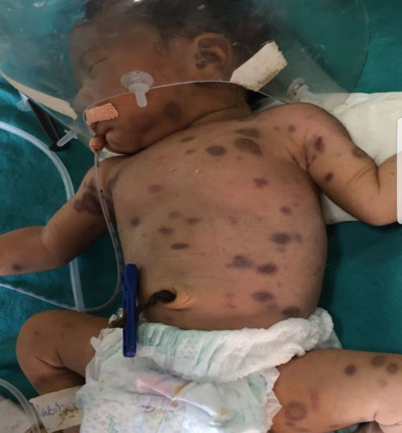
A full-term appropriate for gestational age female
infant born at our hospital (birthweight 3 kg) out of a consanguineous
marriage, to a 27-year-old mother by lower segment cesarian section with
an unremarkable antenatal history, presented at birth with pallor,
palpable purpuric spots and ecchymotic patches, diffuse blueberry muffin
rash of 1 to 3 cm (Fig. 1), and depressed neonatal
reflexes. The craniofacial configuration was normal, but the anterior
fontanelle was full. She had tachypnea and tachycardia. The abdomen was
mildly distended; hepato-splenomegaly was evident on palpation.
 |
|
Fig. 1. Blueberry muffin rash in a newborn with
congenital leukemia and rubella infection.
|
Maternal TORCH profile during the second trimester
done at another hospital was positive for Rubella IgG. Other routine
maternal antenatal investigations, including HbsAg, VDRL, HIV, urine
microscopy, antenatal ultrasound and TSH were normal. Complete blood
count of the child suggested anemia (Hb 5.8 g/dL), thrombocytopenia
(platelet count 14×10 9/L),
leuco-cytosis (total leukocyte count 44.3×10⁹/L)
with blasts 60%, polymorphs 18%, lymphocytes 20% and eosinophils 2%.
Peripheral blood film findings were suggestive of acute leukemia,
showing clumped red blood cells, leucocytosis, blast cells and markedly
reduced platelets, with no hemoparasites.
Blood sugar (85 mg/dL), urea (13 mg/dL), creatinine
(0.4 mg/dL) and bilirubin (total 2.7 mg/dL, indirect 2.1 mg/dL) were
normal. SGOT (331 U/L), SGPT (120 U/L), alkaline phosphatase (1596 U/L),
LDH (6189 IU/L) and GGT (337 U/L) were raised. Uric acid was 4 mg/dl,
calcium was low (7.4 mg/dL), total protein was 5.5 gm/dL (albumin 3.5
gm/dL), and C-reactive protein was non-reactive. INR was raised (2.55),
aPTT was 55 sec, and d-dimer levels were 8.5 µg/mL. Chest X-ray
was normal. Ultrasound abdomen revealed an enlarged liver (7 cm),
contracted gall bladder, and enlarged spleen (6.1 cm). Ultrasound brain
revealed a dilated left lateral ventricle with slight midline shift to
left side, suggestive of intracranial involvement. Skin biopsy was not
done. TORCH profile by Pict Array TORCH ELISA (Nanoplex) was suggestive
of positive Rubella IgM and IgG antibodies.
Flow cytometric immunophenotyping showed 60%
circulating blasts, which expressed CD19, CD20, CD79a, HLA DR, CD34, and
were negative for CD3, CD5, CD7, CD13, CD33, CD117, CD14, cytoplasmic
CD3, and Myeloperoxidase. A diagnosis of B-Cell acute lymphoblastic
leukemia (ALL) with congenital rubella infection was made. Despite
ventilation in neonatal intensive care unit and blood component therapy,
the baby survived only for 3 days.
Discussion
This case fulfilled the criteria for diagnosis of
congenital leukemia: (i) disease presentation at or shortly after
birth, (ii) raised number of immature white blood cells, (iii)
infiltration of cells into extra hemopoietic tissues, (iv)
absence of other conditions like congenital syphilis, blood group
incompatibility, which can cause leukemoid reaction [2].The differential
diagnosis of congenital leukemia includes sepsis and intra-uterine
infections, hemolytic disease of the newborn (HDN) and transient
myeloproliferative disease (TMD) [1]. Infections are ruled out by
serology and culture, while in HDN numerous erythrocyte precursors are
seen in the peripheral smear. TMD is seen usually with Down syndrome,
often with associated transient polycythemia and/or thrombocytosis,
which were not found in this case.
Thrombocytopenia may be seen in up to 80% infants
with CRS, which may be due to decreased megakaryocyte production [6].
Elevated leucocyte counts and severe thrombocytopenia, as seen in this
child, are unusual for rubella. Patients can also develop a hemolytic
anemia that can persist for months and potentially progress to red cell
aplasia with variable leukocyte count [7]. There is paucity of reports
of association of congenital rubella infection with malignancy; one case
of lymphoblastoma has been reported after maternal rubella infection
[8]. Acute myelomonocytic leukemia has also been reported with atypical
congenital rubella [9]. Assembly, maturation and three-dimensional
helical structure of the teratogenic rubella virus using cryo-electron
tomography has also been described [10].
In any newborn presenting with palpable purpura,
besides other causes like common neonatal rashes, leukemoid reactions,
TORCH infection, and bleeding disorder, congenital leukemia should be
considered as a differential diagnosis. On suspicion, immuno-phenotyping
by flow cytometry, together with morphology and cytochemical stains
should be performed. Association of ALL with rubella in this case
suggest that the virus itself may have the potential to cause congenital
leukemia.
Acknowledgment: Dr Priyansha Mathur, Assistant
Professor in Pediatrics for technical help and writing assistance.
Contributors: KSY and SG contributed to diagnostic work-up of
patient. DRB and RS supervised patient management. DRB drafted the
manuscript, which was revised by KSY, SG and RS. All authors approved
the final version of manuscript.
Funding: None; Competing interest: None stated.
References
1. Bajwa RP, Skinner R, Windebank KP, Reid MM.
Demographic study of leukaemia presenting within the first 3 months of
life in the Northern Health Region of England. J Clin Pathol.
2004;57:186-8.
2. Prakash KP, Rau AT, Prakash KP, Rau AT, Bhat ST,
Rau AR, et al. Congenital leukemia – A diagnostic dilemma. Indian
J Med Paediatr Oncol. 2008;29:41-3.
3. Resnick KS, Brod BB. Leukemia cutis in
congenital leukemia: Analysis and review of the world literature with
report of an additional case. Arch Dermatol. 1993;129:1301-6.
4. Mehta V, Balachandran C, Lonikar V. Blueberry
muffin baby: A pictoral differential diagnosis. Dermatol Online
J. 2008;14:8.
5. Li FP, Bader JL. Oncology-epidemiology of selected
childhood cancers. In: Nathan DG, Oski FA, eds.
Haematology of Infancy and Childhood: Philadelphia: WB Saunders,
1987:p.918-41.
6. Hohlfeld P, Forestier F, Kaplan C, Tissot JD,
Daffos F. Fetal thrombocytopenia: A retrospective survey of 5,194 fetal
blood samplings. Blood. 1994;84:1851-6.
7. Franco SA, Riley HD, Jr, Chitwood LA.The
congenital rubella syndrome. South Med J.1970;63:825-30.
8. Stewart A, Webb J, Hewitt D. A survey of childhood
malignancies. Br Med J. 1958;1:1497-508.
9. Kelly SJ, Gibbs T, Cheetham CH. Acute
myelomonocytic leukaemia following atypical congenital rubella. J Clin
Pathol. 1993;46:764-5.
10. Mangala Prasad V, Klose T, Rossmann MG.
Assembly, maturation and three dimensional helical structure of the
teratogenic rubella virus. PLoS Pathog. 2017; 13:e1006377.
|
|
|
 |
|
