|
|
|
Indian Pediatr 2016;53:
42-44 |
 |
Delayed
Cutaneous Findings of Hand, Foot, and Mouth Disease
|
|
Shankha Subhra Nag, Abhijit Dutta and *Rajesh Kumar
Mandal
From Departments of Pediatric Medicine, and
*Dermatology, Venereology, and Leprosy, North Bengal Medical College,
West Bengal, India.
Correspondence to: Dr Shankha Subhra Nag, Department
of Pediatric Medicine, North Bengal Medical College, Sushruta Nagar,
West Bengal, India.
Email: dr.ssnag@gmail.com
Received: February 26, 2015;
Initial review: April 27, 2015;
Accepted: September 30, 2015.
|
Objective: To describe various delayed cutaneous findings
associated with hand, foot, and mouth disease (HFMD).
Methods: Patients presenting with clinical
features of HFMD were followed-up prospectively for a period of 3 months
for the occurrence of delayed cutaneous manifestations.
Results: Out of 68 patients on regular follow-up,
23 (33.8%) showed different types of skin and nail changes following
HFMD. Nineteen showed features of onychomadesis, 9 developed nail
discoloration, and Beau’s line was noted in 5 patients. Cutaneous
desquamation was seen in 7 patients. Spontaneous re-growth of nails
occurred in all cases within 12 weeks follow-up. Skin desquamation
subsided by 2-4 weeks.
Conclusion: Delayed cutaneous findings following
HFMD are common.
Keywords: Beau’s line, Coxsackievirus, Nail discoloration,
Onychomadesis.
|
|
H
and, foot, and mouth disease (HFMD) is a
self-limiting viral infection primarily affecting children under 10
years of age. Human enterovirus 71 and several strains of Coxsackievirus
are causative agents. Clinical features of HFMD are characterized by
erythematous papulo-vesicular eruptions mostly over palms, soles, knees,
buttocks, elbows, and oral mucosa, and may be accompanied with pain and
mild pruritus. Several outbreaks of this disease have been described
from various parts of India since 2005 [1-7].
Delayed cutaneous manifestations following HFMD may
be seen in the form of Beau’s lines, separation of nail plate from nail
matrix (onychomadesis), and desquamation. The present study describes
various delayed cutaneous findings of HFMD following an outbreak in
Siliguri, West Bengal, India.
Methods
This descriptive follow-up study was carried out at
Pediatrics Outpatient Department of the North Bengal Medical College,
West Bengal, India, from July 2014 to December 2014. Clearance from
Institutional Ethical Committee was taken. All children (age 6 months to
15 years) presenting with clinical features of HFMD were enrolled in the
study. Those with recent history of any illness like streptococcal
infection, measles, and Kawasaki disease and those with intake of drugs
implicated in nail matrix arrest (e.g. cloxacillin, valproic acid,
carbamazepine); or trauma to nails were excluded. Patients with atypical
clinical presentation or with possibility of any other diagnoses were
also excluded. Complement fixation test for a panel of antibodies
against coxsackievirus A2, A4, A7, A9, A10, and A16 was performed for
confirmation of diagnosis in some cases. Patients were advised to
follow-up after a week and subsequently every 2 weeks for a period of 3
months. Detailed examination of the skin and nails was performed during
the follow-up visits.
Results
Out of 87 patients registered during the study
period, 19 were lost to follow-up. Among the rest, 23 (12 males)
developed delayed cutaneous findings. Details of initial presentations
are given in Table I. Out of 11 patients who had
serological work-up, 6 showed positive results for coxsackie virus A16
antibody. Nail changes included onychomadesis (19, 82.6%), discoloration
(9, 39.1%), and Beau’s line (5, 21.7%) (Fig. 1 and
Web Fig. 1). More than one finding was observed
in 12 patients. Finger nails were more commonly involved than toe nails.
Number of involved nails ranged from 3 to 20 (mean 13). Onychamadesis
was most commonly observed in nails of middle finger (14, 73.7%),
followed by thumb (12, 63.2%) and ring finger (10, 52.6%). The interval
between appearance of rashes and onset of nail changes range from 17 to
46 (mean 32) days. Nail discoloration commonly involved middle finger
(7, 77.8%), ring finger (5, 55.6%) and little finger (2, 22.2%). It was
diffuse in nature, started proximally and slowly extended distally. In
some cases, lateral nail plate was also involved. Recovery occurred by
spontaneous re-growth of nails within 12 weeks, without any treatment.
TABLE I Initial Presentation in Children With Hand Foot Mouth Disease
|
Symptoms |
No. (%) |
|
Fever |
14 (60.8) |
|
Constitutional symptoms |
07 (30.4) |
|
Itching |
09 (39.1) |
|
Pain/burning sensation |
17 (73.9) |
|
Rashes |
|
|
Palm |
16 (69.6) |
|
Elbow |
21 (91.3) |
|
Other areas in upper limb |
08 (34.8) |
|
Sole |
16 (69.6) |
|
Buttock |
23 (100) |
|
Other areas in lower limb |
08 (34.8) |
|
Genitalia |
03 (13.0) |
|
Trunk |
09 (39.1) |
|
Face |
07 (30.4) |
|
Oral |
15 (65.2) |
 |
|
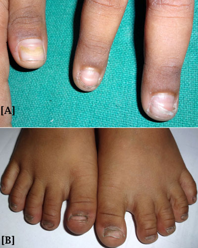
Fig. 1 (a) Onychomadesis, Beau’s line,
and yellowish discoloration of finger nails; (b) Onychomadesis
with loss of the right great toe nail and separation of nail
plate from nail matrix and bed in the left great toe nail.
|
Desquamation limited to periungual and palmo-planter
regions was noted in 7 patients (30.4%) after 2 weeks of follow-up. This
subsided gradually in next 2-4 weeks following topical application of
emollients.
Discussion
Onychomadesis is a non-inflammatory condition
characterized by proximal separation of nail plate from nail matrix with
or without subsequent complete shedding of nails. Besides Coxsackievirus
infection, it may also be seen in streptococcal infection, measles,
Kawasaki disease, epidermolysis bullosa, periungual dermatitis, nail
trauma, and some drugs.
In this study, nail abnormalities were evident in
nearly one-third of the patients of HFMD on follow-up. We also observed
nail discoloration in some patients which has not been described
previously. Several reports [5,8-10] have described nail changes
following HFMD since the temporal relation was first reported by
Clementz, et al. [10]. The mechanism of nail changes still
remains unexplained but it has been proposed that onychomadesis is
caused by inflammation close to the nail matrix [12]. In the outbreak of
HFMD in Finland in 2008, Osterback, et al. [13] detected
Coxsackievirus A6 in shed nail fragments of a patient who had
onychomadesis following a HFMD episode. The authors suggested that
Coxsackievirus A6 replication damages the nail matrix, resulting in
onychomadesis [12]. Nail abnormalities have only rarely been documented
from previous HFMD outbreaks in India [5].
The present study had limitations of a hospital-based
study, a high loss to follow-up, and lack of etiological work-up in
majority of the cases. Moreover, we could not analyze any risk factor or
possible mechanism of these findings.
We conclude that delayed cutaneous findings following
HFMD are common. Parents should be counseled regarding possibility of
these dermatological manifestations and their benign course.
Contributors: SSN: conception of the
study, acquisition of data and drafting the manuscript; AD: design of
the study, acquisition of data and revising the manuscript for important
intellectual content; RKM: analysis and interpretation of data and
revising the manuscript for important intellectual content. All the
authors approved the final version of manuscript.
Funding: None; Competing interest: None
stated.
|
What This Study Adds?
• Delayed cutaneous findings are seen in
about one-third of children with of Hand, foot, and mouth
disease.
|
References
1. Sasidharan CK, Sugathan P, Agarwal R, Khare S, Lal
S, Jayaram Paniker CK. Hand-foot-and-mouth disease in Calicut. Indian J
Pediatr. 2005;72:17-21.
2. Sarma N, Sarkar A, Mukherjee A, Ghosh A, Dhar S,
Malakar R. Epidemic of hand, foot and mouth disease in West Bengal,
India in August, 2007: A multicentric study. Indian J Dermatol.
2009;54:26-30.
3. Arora S, Arora G, Tewari V. Hand foot and mouth
disease: emerging epidemics. Indian J Dermatol Venereol Leprol.
2008;74:503-5.
4. Ghosh SK, Bandyopadhyay D, Ghosh A, Dutta A,
Biswas S, Mandal RK, et al. Mucocutaneous features of hand, foot,
and mouth disease: A reappraisal from an outbreak in the city of
Kolkata. Indian J Dermatol Venereol Leprol. 2010;76:564-6.
5. Kar BR, Dwibedi B, Kar SK. An outbreak of hand,
foot and mouth disease in Bhubaneswar, Odisha. Indian Pediatr.
2013;50:139-42.
6. Kashyap S, Verma GK. Hand-foot-mouth disease:
Outbreak in Shimla. Indian Pediatr. 2014;51:155.
7. Vijayaraghavan PM, Chandy S, Selvaraj K, Pulimood
S, Abraham AM. Virological investigation of hand, foot, and mouth
disease in a tertiary care center in South India. J Glob Infect Dis.
2012;4:153-61.
8. Shikuma E, Endo Y, Fujisawa A, Tanioka M, Miyachi
Y. Onychomadesis developed only on the nails having cutaneous lesions of
severe hand-foot-mouth disease. Case Rep Dermatol Med. 2011;2011:324193.
9. Wei SH, Huang YP, Liu MC, Tsou TP, Lin HC, Lin TL,
et al. An outbreak of coxsackievirus A6 hand, foot, and mouth
disease associated with onychomadesis in Taiwan, 2010. BMC Infect Dis.
2011;11:346.
10. Shin JY, Cho BK, Park HJ. A Clinical study of
nail changes occurring secondary to handfoot-mouth disease:
Onychomadesis and Beau’s lines. Ann Dermatol. 2014;26:280-3.
11. Clementz GC, Mancini AJ. Nail matrix arrest
following hand-foot-mouth disease: A report of five children. Pediatr
Dermatol. 2000;17:7-11.
12. Haneke E. Onychomadesis and hand, foot and mouth
disease–is there a connection? Euro Surveill. 2010;15:pii=19664.
Available from: http://www.eurosur veillance.org/vienAstide.aspx?Articleid=19664.
Accessed December 07, 2015.
13. Osterback R, Vuorinen T, Linna M, Susi P, Hyypia
T, Waris M. Coxsackievirus A6 and hand, foot, and mouth disease,
Finland. Emerg Infect Dis. 2009;15:1485-8.
14. Bracho MA, González-Candelas F, Valero A, Córdoba
J, Salazar A. Enterovirus co-infections and onychomadesis after hand,
foot, and mouth disease, Spain, 2008. Emerg Infect Dis. 2011;17:2223-31.
|
|
|
 |
|
