|
|
|
Indian Pediatr 2014;51: 84 |
 |
Blue Nevus on the Scalp
|
|
Kabir Sardana and *Vivek Sagar
From Departments of Dermatology, Maulana Azad Medical
College, New Delhi, India. and
*ESI Model Hospital, Gurgaon, Haryana, India.
Email: [email protected]
|
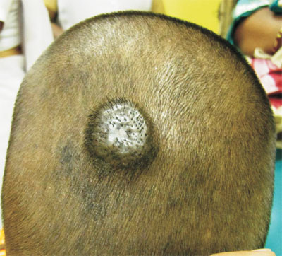
A 6-year-old boy was referred for a firm
3 cm x 2 cm size smooth-surfaced, dome-shaped blue nodule on
the scalp for three years. A possibility of pigmented
histiocytoma/dermatofibroma, pilomatricoma, ossifying
fibroma or blue nevus was kept. Histology revealed a nodular
collection of poorly melanized spindled melanocytes and
deeply pigmented dendritic melanocytes within thickened
collagen bundles which was consistent with a diagnosis of
common blue nevus.
 |
|
Fig. 1 Firm, blue nodule,
with a smooth surface and prominence of follicular
openings.
|
The differentials of a blue nodule
include cavernous haemangioma (soft and large),
dermatofibroma (firm, small and painful), angiokeratoma
(soft and bleeds), blue rubber bleb navus (multiple and
painful), glomus tumour (single, painful) and pilomatricoma
(hard). Blue nevi are rarely seen on the scalp. They are
consequent to a dermal arrest in embryonal migration of
neural crest melanocytes. Larger (>3 cm) lesions warrant a
biopsy to rule out malignant changes. A yearly follow up is
advisable for any recurrence.
|
|
|
 |
|
