|
M
orgagni’s
hernia is a type of congenital diaphragmatic hernia. Affected children are
mostly asymptomatic, diagnosed incidentally by X-ray chest and
confirmed by barium enema. We report a 7 year-old boy who presented with
obstructive jaundice caused by compression of common bile duct (CBD) due
to rotation and stretching of second part of duodenum in a right-sided
Morgagni hernia.
Case Report
A seven-year-old average built phenotypically normal
boy presented with icterus and abdominal pain for 4 weeks. There was no
bleeding diathesis, pruritus or past history suggestive of intestinal
obstruction, recurrent respiratory tract infection or chest trauma. Pain
in the abdomen was colicky and aggravated after food intake; it was
associated with occasional nausea and vomiting.
Clinical examinations revealed mild icterus, normal
growth parameters and stable vitals. Breath sounds were diminished on
right side with overlying dullness on percussion. The shape of the chest
and abdomen, movement with respiration and bowel sounds were normal. Rest
of the systemic examination unremarkable.
Complete blood count, serum electrolytes and renal
function test were within normal limits. Total bilirubin was 6.5 mg/dL),
(conjugated 4.5 mg/dL, ALT 111U/L, AST 141U/L, and high alkaline
phosphatase level. Total protein was 7.3g/dL (albumin 4.3 g/dL).
Prothrombin time was 13 s (control 12.8 s). Serum amylase and lipase level
were within normal limit. Serology for hepatitis A and B (HB SAg,
IgM HAV) were negative. X-ray chest revealed opacities in right
middle and lower zone (Fig.1). Ultrasonography of the chest
detected gut loops which appeared to be entering the right pleural space,
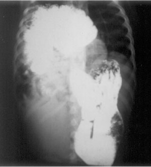
suggesting herniation of gut. Barium meal follow through confirmed right
side diaphragmatic hernia (Fig.2). CT scan of chest and
upper abdomen revealed right-sided Morgagni’s type of diaphragmatic hernia
with features of gastric outlet obstruction and the pancreatic head was
being pulled up superiorly resulting in stretching and compression of
retro-duodenal portion of common bile duct (CBD) and collapse of right
lower lobe of lung and contralateral shift of mediastinum.
Electrocardiogram and 2-dimensional echocardiography revealed no
abnormality.
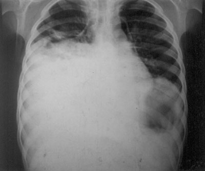 |
| Fig. 1 X-Ray chest showing
opacities in right mid and lower zone
|
 |
|
Fig. 2 Barium meal follow through showing
right sided diaphragmatic hernia. |
He was treated surgically. The abdominal contents of
the thorax were brought down; the CBD obstruction got spontaneously
corrected. Associated malrotation was also corrected. The defect was
repaired with vertical mattress sutures of interrupted 2-0 prolene
stitches. Immediate postoperative period was uneventful. The
child is well on follow-up.
Discussion
The most common type of CDH is Bochdalek hernia (posterolateral
defect) which manifests itself soon after birth. The other type of CDH is
Morgagni hernia (retrosternal hernia); which occurs due to development
defect caused by failure of fusion between the fibrotendinous elements of
sternal and costal portion of the diaphragm. The symptoms of Morgagni
hernia usually do not become apparent until attainment of adulthood,
usually after 50 years of age. In younger age group it occurs
predominantly in males; however, among the elderly it predominates in
female. This hernia is rare in children, representing only 1%-6% of all
types of CDH [1]. The reason behind rarity among the children may be that
increased intra-abdominal pressure with advancing age is require to
stretch the defect and cause herniation of abdominal content in to thorax
[2]. The diagnosis of this hernia is often delayed because most of the
patients are asymptomatic; presence of hernia is detected only
incidentally on chest X-ray [3]. If symptomatic, it produces
variable nonspecific respiratory or gastrointestinal symptoms but rarely
acute intestinal obstruction and colonic perforation as presenting
features have been reported [4].
The transverse colon or omentum is the usual content of
this hernia; stomach or portions of the liver are rare. Obstruction of the
extrahepatic biliary system by the herniated content is thus rare.
Caldeiro, et al. [5] coined the term "choledochal semivolvulus" to
describe these findings. Obstructions of the CBD due to involvement into a
hernia sac, as well as traction and volvulus formation of the CBD, were
common explanations of such biliary obstruction [6].
The diagnosis of Morgagni’s hernia is usually
established by chest X-ray with a lateral film to show the
anteriorly placed bowel loops. This can be further confirmed by barium
enema or by barium meal and follow-through because colon is usually the
most common content of the hernial sac. The diagnosis can sometimes be
difficult if the hernial sac contains omentum or liver [7]. USG, CT scan
or MRI thorax is advocated in such a situation [3].
After the diagnosis of Morgagni’s hernia, all cases
should be operated to avoid the risk of bowel strangulation and
perforation. We performed an open transabdominal repair of the
diaphragmatic defect. Other treatment options include laparoscopic surgery
[8] and video-assisted thoracic surgery [9].
Acknowledgment: Prof BK Chatterjee, Head, General
Surgery; Dr P Deb (Radio-diagnosis) and Dr D Roy (Anatomy) for their
support.
Contributors: MR: concept and design; AKB:
supervised the patient diagnosis and management and revised the
manuscript; SB: operative management, manuscript preparation; TP: patient
diagnosis and management, literature search.
Funding: None.
Competing interests: None stated.
References
1. Cullen ML, Klein MD, Philippart AI. Congenital
diaphragmatic hernia. Surg Clin North Am. 1985;65: 1135-8.
2. Loong TPF, Kocher HM. Clinical presentation and
operative repair of hernia of Morgagni. Postgrad Med J. 2005;81:41-4.
3. Minneci PC, Deans KJ, Kim P, Mathisen DJ. Foramen of
Morgagni hernia: changes in diagnosis and treatment. Ann Thorac Surg.
2004;77:1956-9.
4. Cakmak O, Pektas O, Baskin D. Retrosternal hernia (Morgagni)
with colonic perforation due to incarceration. Pediatr Surg Int.
1990;5:274-5.
5. Caldeiro JC, Curcio A, Gigena VC, Barbarosa G.
Choledochal semi volvulus with jaundice due to hiatal hernia. Initial
percutaneous management. Acta Gastroenterol Latinoam. 2001;31:329-32.
6. Glielmi M. Unusual case of cholestatic jaundice
caused by gigantic Morgagni-Larrey hernia in childhood. Acta Chir Ital.
1963;19:1381-90.
7. Groff DB. Diagnosis of a Morgagni hernia complicated
by a previous normal chest X-ray. J Pediatr Surg. 1990;25:556-7.
8. Kuster GG, Kline LE, Garzo G. Diaphragmatic hernia
through the foramen of Morgagni: laparoscopic repair: case report. J
Laparoendosc Surg. 1992; 2: 93-100.
9. Hussong RL, Landreneau RJ, Cole FH. Diagnosis
and repair of a Morgagni hernia with video assisted thoracic Surgery. Ann
Thorac Surg. 1997;63:1474-5.
|


