Polyarteritis nodosa (PAN) is a rare multisystem
disease of childhood characterized by necrotising vasculitis affecting
medium sized arteries(1). We present a case of PAN with an unusual
association i.e., stenosis of a main renal artery.
Case Report
A three-year-old boy, first born to
non-consanguineous parents, previously in good health, developed fever
which was insidious in onset, low grade, intermittent, for a period of 3
months. The fever was associated with generalized muscle pain and
painful swelling of knees and ankles on both sides, severe enough to
prevent the boy from walking. This was followed by blackish
discoloration of the skin overlying the same joints as well as the tip
of fingers in both the hands. He had malaise and significant weight
loss. There was no history of hematuria or oliguria.
On examination, the boy was conscious and afebrile.
Dry gangrene involving the distal ends of the fourth and fifth finger on
the left and the third finger on the right was noted. The skin overlying
the anterior aspect of both the knees showed scarring and punched out
ulcers. Multiple fine violaceous nodules were noted in both the palms.
All the peripheral pulses were well felt and the BP was 120/70 mmHg
(<95th percentile). There was no renal bruit.
Investigations revealed the following: Hb 12 g/dL, TC
12,900 cells/cu.mm, DC N 63%, L 37%, ESR 110 mm in one hr and platelet
count 6 lakhs/cumm. Serum HBsAg, antineutrophil cystoplasmic antibody (ANCA)
and antistreptolysin O (ASLO) titer were negative. Liver enzymes were
within normal limits. Urine analysis and renal function tests were
normal. Coagulation profile including antiphospholipid antibody did not
reveal any abnormality. Ultrasonography of the abdomen and
echocardiogram were normal.
In view of the fever, severe myalgia, weight loss,
the characteristic gangrenous changes and modular lesions with no
evidence of glomerulonephritis the boy was diagnosed to have classical
PAN. He was treated with oral prednisolone (2 mg/kg/day) for 8 weeks. As
he did not respond to steroids, IV cyclophosphamide (500 mg/m2) was
given once in 4 weeks for 6 months in addition to oral prednisolone.
Thereafter he was advised oral prednisolone (0.5 mg/kg on alternate day)
for 2 years. In addition, acetyl salicylic acid (5 mg/kg/day) was
prescribed in view of thrombocytosis and prevention of further
thromboembolic complications.
However two years later, the boy presented again with
polyarthritis, fever, myalgia, abdominal pain, malena and gangrenous
change of the skin overlying the right ankle. History revealed that he
had stopped all drugs 6 months prior to admission. On examination, his
blood pressure was found to be high (140/100 mmHg) (>95th percentile)
with serial measure-ments. Investigations revealed the following: Hb
12.5 g/dL, TC 24,000 cells/cu.mm, DC N 70%, L 27%, E 3%, ESR 90 mm in
one hr and platelet count 7.4 lakhs/cu.mm. Bleeding time, clotting time,
prothrombin time and partial thromboplastin time were within normal
limits. Renal function tests were also normal. A flush abdominal
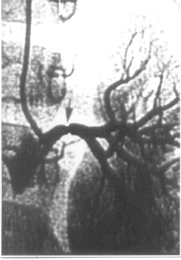
aortogram and bilateral selective renal angiography was done which
revealed moderate stenosis of the left main renal artery (Fig.1).
However, there were no aneurysms or segmental narrowing of the intra
renal vasculature. In view of the severe abdominal pain and malena, IV
methyl-prednisolone (30 mg/kg/day) was given for 7 days. The
hypertension was controlled with enalapril (0.25 mg/kg/day). At
discharge, he was relieved of his symptoms and had a BP of 120/80 mmHg.
However, the child was subsequently lost for follow up.
 |
|
Fig. 1. Angiography showing stenosis of left
renal artery. |
Discussion
The initial diagnosis of PAN is clinical. Biopsy of
the involved tissue like skin, muscle, sural nerve or kidney may support
the diagnosis. Angiography findings include aneurysms, segmental
narrowing and variations in the caliber of arteries, together with
pruning of peripheral vascular tree often found in the kidney and or
liver(2,3). According to Brogan, et al.(4) the most reliable non
aneurysmal signs in angiography are perfusion defects, presence of
collateral arteries, lack of crossing of small peripheral arteries and
delayed emptying of small renal arteries. Both PAN and Takayasu
arteritis may involve the renal arteries and present with severe
hypertension due to renal artery stenosis(5). However, arteritis limited
to the main renal artery with no involvement of intrarenal vasculature
as found in our case is an unusual variant of PAN. This renal artery
stenosis may be due to the arteritis(6) or just a coexistent finding and
probably contributing to the hypertension. The very young age of onset
of PAN in this boy also makes the case interesting. Progressive renal
insufficiency can occur during the acute course of classical PAN due to
renal vascular involvement without glomerulonephritis(7). Hence, in a
case of PAN with severe hypertension, the exact cause for the
hypertension should be ascertained and not presumed to be caused by
intrarenal aneurysms.
Contributors: BA and TV collected the data. BRN
drafted the manuscript. MVK reviewed the manuscript and will act as a
guarantor for the paper.
Funding: None.
Competing interests: None stated.
