|
A one and a half-year-old girl was brought to
us with a slowly growing abdominal mass, first noticed by her
parents at 2 months of age. There were no bowel or urinary
complaints. Abdominal examination revealed a firm, nontender,
fixed, transversely placed mass measuring 20×15 cms. Laboratory
test findings were unremarkable. A plain radiograph revealed a
large, partially calcified soft tissue mass in the right side of
the abdomen with long bone-like structures traversing the middle
of the abdomen. Ultrasonography revealed a large, hyper-echoic,
intra-abdominal mass with areas of calcifications. The MRI scan
showed a large mass having a round head like structure in the
right sub-hepatic space. The vertebral axis was visualized in its
entire length going from the right upper abdomen to the left lower
abdomen (Fig. 1). Multiple long bone-like structures were
also identified. Based on the MRI findings, a preoperative
diagnosis of fetus-in-fetu was made. The patient underwent
laparotomy and complete excision of the mass. The radiograph of
the specimen showed few calcified long bones with no identifiable
vertebral bodies. The pathological examination revealed a large
mass composed of adipose tissue and covered with skin. The mass
also had evidence of bone formation. Vertebral bodies could be
identified along the length of the fetiform mass with bone marrow
formation. Sections taken between the vertebral bodies
demonstrated formation of neural tissues.
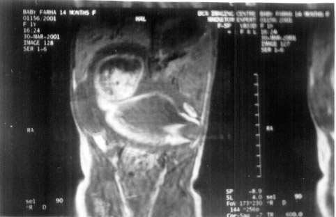 |
| Fig.1. MRI scan
demonstrating the cranial ring in the right subhepatic
space along with the vertebral axis in its entire length |
Fetus-in-fetu is a term used to describe the
inclusion of one fetus within the body of another. Fewer than 100
cases have been reported in litertature till date. It is
controversial whether fetus-in-fetu is a distinct entity or
represents a highly organized teratoma. The identification of a
vertebral column suggests the diagnosis of fetus-in-fetu rather
than teratoma. Occasionally, an underdeveloped and markedly
dysplastic spinal column prevents the identification of the
vertebral bodies at imaging(1). A recent review on the subject
reported that a preoperative diagnosis of fetus-in-fetu was made
in only 17.67% of all the cases till 1980 and the vertebral column
was not identified in about 9% of cases even after the
pathological examination(2). Though CT scan has proved very
helpful in preoperative diagnosis, the non-visualization of the
vertebral column on CT scan does not exclude this diagnosis(2).
Although Magnetic Resonance Imaging (MRI) seems to be an ideal
technique for demonstrating the wide range of tissue within such
lesions, there are only a few reports of the use of MRI in the
identification of the lesion(2,3). MRI allows imaging in the
sagittal and coronal planes and does not rely on calcification for
demonstrating tissues. This helps in identifying insufficiently
calcified vertebrae and vertebral axis. In our case, the vertebral
axis was demonstrated along the whole length on the coronal
sections of the MRI. There was an excellent delineation of the
tissues on MRI with good delineation of the cranial ring
suggestive of the fetal skull which was not picked up on the
conventional radographs including specimen radiographs.
We propose that MRI has the potential as imaging modality of
choice for the diagnosis of fetus-in-fetu.
A. Sinha,
Y.K. Sarin,
M. Sengar,
Department of Pediatrics Surgery,
Maulana Azad Medical College,
New Delhi - 110 002, India.
E-mail: [email protected]
.
1.
Knox JS, Webb AJ. The clinical features and treatment of fetus
in fetu: two case reports and review of literature. J Pediatr
Surg 1975; 10: 483-489.
2. Hoeffel CC,
Nguyen KQ, Tran TT, Fornes P. Fetus in fetu: A case report and
literature review. Pediatrics 2000; 105: 1335-1344.
3. Hanquinet S, Damry N, Heimann
P, Delaet MH, Perlmutter N. Association of a fetus in fetu and
two teratomas: US and MRI. Pediatr Radiol 1997; 27: 336-338.
|
