|
|
Case Reports Indian Pediatrics 2002; 39: 79-83 |
||||
|
Familial Hodgkin’s Disease in Two Siblings |
||||
|
V. Thavaraj Rajive Kumar L.S. Arya
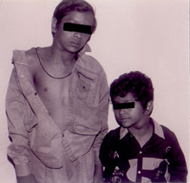
Hodgkin’s disease is a malignant disease usually arising in the lymph nodes. The childhood form of Hodgkin’s disease occurs in patient’s 14 years old or younger(1). Familial Hodgkin’s disease is very rare. We report in this paper two siblings presenting with Hodgkin’s disease at the same time. Case Reports Sibling 1: A 14-year-old male child was apparently well till 4 years ago, when he had fever intermittent in nature lasting for 2-3 days, high grade with no chills or rigors or night sweats. The fever subsided with antipyretics. Then he noticed swelling in the neck which continued to enlarge. He was started on anti-tuberculous drugs on the basis of a positive Mantoux test of 32 mm and chest roentgenogram suggestive of hilar lymph nodes. He received anti tubercular drugs for 9 months. The swelling in the neck persisted and there was no increase in the size of swellings. He remained asymptomatic for 3 years. Then a fresh group of swellings appeared at the base of the neck. There was no fever or other symptoms associated with the swellings. Lymph node biopsy was done at this time and a diagnosis of Hodgkin’s disease was made outside the institute. When he presented to the Pediatric Oncology Clinic at the All India Institute of Medical Sciences, New Delhi he was febrile, the right cervical lymph nodes were 3-4 in number, 2-4 cm in size, firm and rubbery in consistency. The right supraclavicular lymph nodes were also enlarged (Fig. 1 ). There were no axillary or inguinal lymph nodes. The liver and spleen were not palpable. The hemo-globin level was 11.2 g/dl. The total leukocyte count was 9.4 × 10⁹/L. The platelet count was 190 × 109/L. The erythrocyte sedimentation rate was 45 mm in 1st hour by Westergren method. The chest X-ray was normal. There was no mediastinal disease. The contrast enhanced computerized tomography (CECT) of the chest and abdomen showed no evidence of any nodal enlargement in the abdomen, pelvis, mediastinum and hilum. Nodes were seen to be present in the supraclavicular region. The lungs, liver, spleen were free of the disease. He was graded as stage II disease after CECT. The bone marrow biopsy was normocellular with normal hemopoietic cells of all series. There was no evidence of lymphoma deposits. The lymph node biopsy was reviewed by us and it was compatible with Hodgkin’s disease of mixed cellularity type. The IgM mono spot test for Epstein-bar virus (EBV) was non reactive. The IgM antibody titers to cytomegalovirus (CMV), rubella virus and toxoplasma were negative. The IgM antibody titers to human herpes virus (HHV 1&2) was reactive. The immuno-globulin levels were normal and HIV ELISA was negative. Sibling 2: This 7-year-old male child (younger brother of the previous case) presented with a history of swelling in the neck for the last 12 months. The swelling started after a bout of fever which lasted for 4-5 days and subsided with antipyretics. The swelling in the neck was gradually increasing in size. There was no history of anorexia, loss of weight, fever with chills or night sweats, icterus, increasing pallor, and body pains. He was investigated by a private practitioner for tuberculosis. The erythrocyte sedimentation rate was raised at that time. Even though the Mantoux test was non reactive and the fine needle aspiration of lymph nodes showed reactive lyumphadenitis he was started on antitubercular drugs by a private doctor. He received antitubercular drugs [Isoniazid, Rifampicin and Pyrazinamide] for 6 months. The swellings started increasing in size. The lymph node biopsy was carried out which revealed mixed cellularity type Hodgkin’s disease. At the time of presentation to our clinic he was afebrile. There was no pallor, icterus and pedal edema. The lymph nodes in the left cervical region were 2-3 in number, 2-6 cm in size, non-tender, non matted and rubbery in consistency (Fig. 1). No other group of lymph nodes were palpable in the neck, axilla or inguinal region. Liver was not palpable and spleen ws 2.0 cm below the costal margin. Testes were normal. The lymph node biopsy was reviewed by us and was compatible with Hodgkin disease mixed cellularity type.
Fig. 1. Familial Hodgkin’s disease in two siblings aged 14 and 6 years of age. The hemoglobin was 11.0 g/dL, the total leukocyte count was 0.5 × 10⁹/L. The platelet count was 2.64 × 10⁹/L. The differential counts showed an increase in monocytes and eosinophils. The erythrocyte sedimentation rate was 10 mm at 1 hour by Westergren method. The chest X-ray showed prominence in both hilar shadows due to hilar node enlargement. The CECT revealed enlarged lymph nodes in left supraclavicular, carinal, post carinal, right hilar, peri pancreatic and retroperitoneal regions with multiple small focal defects in the spleen. He was in stage III disease. The bone marrow biopsy was normocellular with normal hematopoietic cells. There was no evidence of lymphoma deposits. The IgM antibody to EBV was non reactive. The IgM tests for cytomegalovirus, rubella, toxoplasma and human herpes virus were negative. The immunoglobulin levels were normal and HIV ELISA was negative. The family is a Hindu single unit family hailing from the state of Uttar Pradesh. There was no history of consanguinity. Besides these two brothers there is a sister who is 9 years of age and is healthy. There was no history of Hodgkin’s disease or any other cancer in the family members and in the first degree relatives. There was no history of any member suffering from any illness in the recent past suggestive of infectious mono-nucleosis. Both the brothers were started on chemotherapy protocols. They received four alternating cycles of Cyclophosphamide, Prednisolone, Vincristine, and Procarbazine (COPP) and Adriamycin, Bleomycin, Vinblastin and Dacarbazine (ABVD) regimes. The lymphnode enlargement completely regressed in both the siblings. Discussion Children account for about 10% of the patients with Hodgkin’s disease. Familial Hodgkin’s disease is estimated to represent approximately 1-1.5% of all cases of Hodgkin’s Disease(2). In a review on familial Hodgkin’s disease(3), 22 cases of children with familial disease were documented. The authors reported familial Hodgkin’s disease to represent approximately 4.5% of all cases of Hodgkin’s disease. Shared environmental factors such as EBV and other viral agents and genetic determinants have been proposed to explain familial aggregation of risk of Hodgkin’s disease. In 1959 Razis et al. studied the magnitude of risk of family members developing Hodgkin’s disease. They estimated a 3 fold increased risk of Hodgkin’s disease in first degree relatives(3). Grufferman et al. found a seven fold increased risk for siblings of young adult cases and no increased risk for sibling of older adult cases(4). In this report, the age of the elder sibling was 14 years and the younger one was 7 years. The mean age of the sibling pair was 10 years, which is much lower than the reported cases in literature. The median age of sibling pairs with Hodgkin’s disease reported by Siebert et al was 27 years(5). Fraumeni et al. reported only two sibling pairs between 14 years and 19 years who had Hodgkin’s disease in a retrospective analysis of 359 cases of children with Hodgkin’s disease who had died between 1960-1964 in USA(6). None of the first degree relatives of these two brothers had Hodgkin’s disease. Haim et al had projected a 9 fold increased risk for the Hodgkin’s disease in the first degree relatives of patients with Hodgkin’s disease(7). Cancer risk in the first degree relatives of cancer probands is estimated as 1.27 and 1.68 for Hodgkin’s disease and non Hodgkin’s lymphoma, respectively in a population based estimation(8). Fraumeni et al. had found that out of 314 children with Hodgkin’s disease only 3 confirmed instances of Hodgkin’s disease were found in close relatives(6). The parents of these two siblings were normal without Hodgkin’s disease or any other cancer. The parent sibling pair having Hodgkin’s lymphoma have been described earlier(5). In two generation pairs, Hodgkin’s disease in the parent was always associated with Hodgkin’s disease in the child, whereas non Hodgkin’s lymphoma in the first generation was found in association with both non-Hodgkin’s lymphoma and Hodgkin’s disease in the second generation(5). Horizontal transmission patterns was predominant in sibling pairs with Hodgkin’s disease as compared to vertical transmission in non-Hodgkin’s lymphoma pairs(5). The siblings were of the same sex. The other sister is normal without Hodgkin’s disease. The sex concordance was seen more in pairs with non Hodgkin’s lymphoma than in pairs with Hodgkin’s disease(5). These two siblings presented at the same time with Hodgkin disease. Even though the elder one had symptoms lasting for 3 years. The younger sibling had symptoms for only 12 months. The median time interval between diagnosis in non Hodgkin’s lymphoma pairs was less than 3.5 years, whereas for Hodgkin’s disease there was no significant difference in the time interval between diagnosis among the siblings(5). Several epidemiological studies have suggested that shared exposure to infectious agents including Herpes virus 6 (HHV-6), Herpes Virus 7 (HHV-7), Cytomegalo virus (CMV) and Epstein Barr virus (EBV) may be involved in the clustering of Hodgkin’s disease in families(1). There is evidence of a three fold increased risk of Hodgkin’s disease among people with previous history of infec-tious mononucleosis(9). The IgM antibody to EBV was negative in these two siblings. EBV infection has been proposed to explain familial aggregation(10). Studies on familial Hodgkin’s disease failed to detect high antibody titer to EBV in the affected relatives(11). Genetic susceptibility to Hodgkin’s disease has been studied. In this report even though both the brothers were affected by the same disease, neither the parents nor the sister had developed Hodgkin’s disease. In a study, in 432 sets of twins affected by Hodgkin’s disease in adults before the age of 50 years it was found that monozygotic twins had greatly increased risk, whereas there was no increase in the risk for dizygotic twins of patients with Hodgkin’s disease, thereby suggesting a genetic susceptibility(12). Even though we had not studied HLA antigens in these two brothers, HLA concord-ance among affected pairs of siblings(13) and excess of HLA antigens have been shown in the families at risk(14). In the last 15 years around 250 cases of childhood Hodgkin’s disease have been seen in our clinic. This is the first time we have seen two siblings affected in the same family at the same time. There is no other report of familial Hodgkin’s disease in children in Indian literature. Environmental and genetic factors were looked into and we could not identify any such factor. Contributors: The cases were under the care of VT and LSA who drafted the manuscript. RK was responsible for histopathological description and interpretation. VT will act as the guarantor for the manuscript. Funding: None. Competing interests: None stated.
References
|
|
|
![]()