|
|
|
Indian Pediatr 2020;57: 179 -180 |
 |
Cardiac Rhabdomyoma Causing Progressive Dynamic Severe Right
Ventricular Outflow Tract Obstruction in an Infant
|
|
Amitabh Poonia *,
Priya Giridhara and Arun Gopalakrishnan
Department of Cardiology, Sree Chitra Tirunal
Institute for Medical Sciences and Technology, Thiruvananthapuram,
Kerala, India.
Email: [email protected]
|
|
Multiple cardiac masses were
incidentally detected in a neonate on twelve day of life. Failure to
thrive, feeding difficulty and severe dynamic right ventricular outflow
tract obstruction developed at 7 months of age. Surgical resection of
intracardiac masses relieved symptoms and histological studies confirmed
rhabdomyoma. Progressive increase in the size of rhabdomyoma during
infancy is an uncommon presentation and surgery can be life-saving.
Keywords: Cardiac tumor, Echocardiography,
Tuberous sclerosis.
|
|
A
12-day-old asymptomatic neonate was detected to
have multiple cardiac masses during evaluation of a cardiac murmur.
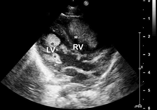
There were multiple lobulated cardiac masses in the left ventricular
apical region and interventricular septum (largest 10 x 8 mm) and
pedunculated mass (14 × 9 mm) in right ventricular outflow tract (RVOT)
(Fig. 1). The baby did not have other features of tuberous
sclerosis and was kept on close medical follow-up.
 |
|
Fig. 1 Echocardiogr am (parasternal
long-axis view) showing multiple cardiac masss in left ventricle
(LV) and right ventricle (RV).
|
At 7 months of life, parents reported failure to
thrive and new onset feeding difficulty. The right ventricular mass had
increased in size (19×19 mm) and was causing severe RVOT obstruction
(peak gradient 86 mmHg) without increase in size of left ventricular
masses. In view of symptomatic severe RVOT obstruction, surgical
resection of all the masses was done. The largest mass (20×15 mm) was
firm in consistency, gray-white and glistening, arising from right
ventricular free wall partly attached to the chordae of septal leaflet
of tricuspid valve (Web Fig. 1a).
Histopathology showed vacuolated tumor cells with clear cytoplasm and
characteristic spider cells on Haematoxylin and Eosin staining (Web
Fig. 1b) and Desmin expression (Web Fig. 1c)
suggestive of cardiac rhabdomyoma.
Neonatal cardiac tumours are rare, rhabdomyomas being
commonest among them. Tuberous sclerosis is associated with cardiac
rhabdomyomas in 50-60% patients and conversely, rhabomyomas are
associated with tuberous sclerosis in 59-80% [1,2].
Rhabdomyomas are generally multiple,
well-circumscribed, intramural or pedunculated tumours seen most
commonly in the ventricles. They are hamartomas with no malignant
potential. Their presentation varies from asymptomatic incidentally
detected cardiac murmur, congestive cardiac failure, arrhythmias or
sudden infant death depending on the size, number and location of the
tumour.
Cardiac rhabdomyomas have a propensity for
spontaneous regression [3,4]. Most of them have a benign course and
remain static or regress with age, higher chances of spontaneous
regression seen at younger age. Complete regression is common in the
ûrst 4 years of life [3,4].
Mammalian targets for rapamycin inhibitors have been used to treat
large, inoperable or residual rhabdomyomas [5]. Surgical intervention is
indicated with haemodynamic compromise or intractable arrhythmia.
Progressive severe dynamic outflow tract obstruction is an uncommon
presentation and surgery can be life-saving.
Contributors: AP,PG: prepared the manuscript; AG:
edited the manuscript. All authors have approved the final version.
Funding: None; Competing interest: None
stated.
References
1. Ajay V, Singhal V, Venkateshwarlu V, Rajesh SM.
Tuberous sclerosis with rhabdomyoma. Indian J Hum Genet. 2013;19:93-5.
2. Harding CO, Pagon RA. Incidence of tuberous
sclerosis in patients with cardiac rhabdomyoma. Am J Med Genet
1990;37:443-6.
3. Farooki ZQ, Ross RD, Paridon SM, Humes RA,
Karpawich PP, Pinsky WW. Spontaneous regression of cardiac rhabdomyoma.
Am J Cardiol. 1991;67:897-9.
4. Bosi G, Lintermans JP, Pellegrino PA,
Svaluto-Moreolo G, Vliers A. The natural history of cardiac rhabdomyoma
with and without tuberous sclerosis. Acta Paediatr 1996;85:928-31.
5. Breathnach C, Pear J, Franklin O, Webb D, McMahon
CJ. Rapid regression of left ventricular outflow tract rhabdomyoma after
sirolimus therapy. Pediatrics. 2014;134:1199-202.
|
|
|
 |
|
