|
|
|
Indian Pediatr 2011;48: 141-143 |
 |
Ultrasonography for Masseter Muscle
Cysticercosis |
|
Anjali Gokarn, Suhas Gokarn and Vivek Rathod
From Vikas Children’s Hospital, Vasai, Maharashtra,
India.
Correspondence to: Dr Anjali Gokarn, Asmita, Phadke Wadi,
Vasalai, Vasai 401 201, Maharashtra.
Email; [email protected]
Received: April 27, 2009;
Initial review: May 12, 2009;
Accepted: September 4, 2009.
|
Solitary cheek swellings can present a diagnostic dilemma. We managed
two children 10 y and 8 y presenting with pain and swelling on one side
of cheek for over 15 d and no constitutional symptoms. Sonography showed
cysticercosis in both of them. We treated both with steroids and
albendazole, with good response.
Key words: Cysticercosis, Masseter muscle, Ultrasonography.
|
|
Cysticercosis,
the infestation with the encysted larval stage of the parasite T.
Solium commonly infests the brain, but muscles are also often affected
[1-6]. Intramuscular cysticercosis has non-specific mani-festations and
diagnosis can be difficult. High resolution sonography (USG) can
demonstrate the classical cyst with scolex within, and is a convenient
test for diagnosis [1-3]. We present two patients with solitary cheek
swellings where USG helped diagnose masseter muscle cysticercosis.
Case Reports
Case 1: A 10 year old girl, resident of Mumbai, was
brought with a painful swelling over the right cheek for 2 months. There
was no fever or other symptoms. The whole right cheek looked swollen and
on palpation the swelling was tender, globular, 3 cm in diameter and felt
firm in the center. We suspected a hematoma or soft tissue tumor. Blood
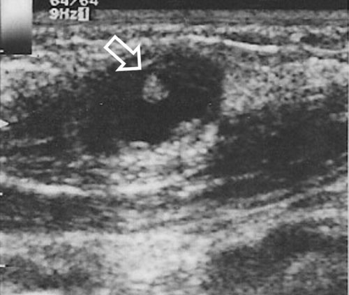
counts were normal. High resolution ultrasonography of the swelling (Fig.
1) revealed a well defined cystic mass with an eccentric echogenic
nidus, the scolex, (arrow) within the masseter muscle fibers. There was
surrounding edema fluid collection. An MRI of the swelling showed similar
findings. We treated her with oral prednisolone 2mg/kg for 4 days and
albendazole 15mg/kg for 28 days. The swelling disappeared after treatment.
 |
|
Fig.1
Ultrasonography
of masseter showing a cystic mass and an eccentric echogenic nidus
(arrow) suggestive of scolex. |
Case 2: An 8 year old girl, resident of Nepal,
presented with a similar, painless swelling over the left cheek for 15
days. There were no constitutional symptoms and blood counts were normal.
Clinically, the swelling was 2 cm in diameter, firm, tender and mobile.
High resolution ultrasonography revealed a 1.1 cm cystic mass with an
eccentric scolex. There was no perilesional edema. We did not do a MRI. We
treated her with steroids and albendazole. The swelling reduced after
treatment and repeat sonography showed reduced size of cyst and absence of
the scolex.
Discussion
Diagnosis of intramuscular cysticercosis is difficult
solely on a clinical basis as the manifestations are not specific and
lesions may be confused with lipoma, fibroma, neurofibroma or
intramuscular abscess [4]. Serological diagnosis using Elisa has >90%
sensitivity and specificity and is positive in persons with many
parenchymal cysts. However cases with solitary lesions (like both our
patients) or old calcified disease may not have detectable antibodies [7].
Fine needle aspiration cytology has been extensively used for diagnosis of
intramuscular cysticercosis. However, it is a invasive test and a blind
procedure and in some cases the aspirated smears are non-specific [8].
This test has low sensitivity, is invasive, expensive and time consuming.
Plain radiography rarely shows cysticerci in the active
phase, but show calcified lesions in chronic cases [2]. Calcified
intramuscular cysticerci appear as millet shaped elliptical lesions in the
soft tissue parallel to muscle fibres [4]. Plain X-ray in a patient
with a solitary cyst has a poor yield. MRI is extensively used for
diagnosing neurocysticercosis where it can clearly show the cyst with the
scolex within [7].
High resolution sonography provides all information
available with MRI, and more with regards to muscle pathology [9]. The
diagnostic feature of a cysticercus granuloma is the presence of an oval
or rounded well defined hypoechoic cystic lesion with smooth walls and an
eccentric hyperechoic nidus representing the scolex within [1]. The cyst,
particularly the scolex, may be better visualized by USG than MRI, in
muscular lesions [4]. The radiological appearances and clinical features
correlate with the stage of maturation of the disease. When the parasite
is alive, in the initial stage of the disease, the cyst is small, without
perimeter enhancement, as seen in our second patient. The patient may or
may not be symptomatic [4]. In later stages, a homogenous hypoechoeic soft
tissue lesion around the characteristic cyst corresponds to leakage of
fluid on death of the parasite [1]. This elicits an intense inflammatory
response in the tissues. This may at times be mistaken for an
intramuscular abscess, but the characteristic cyst with scolex clinches
the diagnosis. The patient is symptomatic at this stage and may have
waxing and waning swelling [4]. Our first patient had similar clinical and
USG findings.
At times the scolex within the cyst may not be seen and
the cyst may appear irregular with minimal fluid on one side indicating a
leakage of fluid. It may be due to escape of the scolex to outside the
cyst [1]. A careful search in the inflammatory fluid for the scolex, will
be fruitful. An elliptical calcified lesion in the muscle along the muscle
fiber is the final stage. The patient is usually asymptomatic at this
stage [4].
USG done in both patients had clearly shown the cyst
with its characteristic scolex. MRI done in the first patient did not add
new information, so we did not do this expensive test in the second child.
Both of them responded well to conservative treatment.
Contributors: AG and VR were involved in patient
management. SG prepared the manuscript and revised it. AG reviewed the
literature and will act as a guarantor.
Funding: None.
Competing interests: None stated.
References
1. Vijayraghavan S. Sonographic appearances in
cysticercosis. J Ultrasound Med. 2004;23:423-7.
2. Asrani A, Morani A. Primary sonographic diagnosis of
disseminated muscular cysticercosis. J Ultrasound Med. 2004;23:1245-8.
3. Mittal A, Das D, Iyer N, Nagaraj J, Gupta M.
Massetter cysticercosis – a rare case diagnosed on ultrasound.
Dentomaxillofacial Radiol. 2008;37:113-6.
4. Jankharia B, Chavan G, Krishnan, Jankharia B. MRI
and Ultrasound in solitary muscular and soft tissue cysticercosis.
Skeletal Radiol. 2005;34:722-6.
5. Kumar A, Bhagwani D, Sharma R, Kavita, Sharma S,
Datar S. Disseminated cysticercosis. Indian Pediatr. 1996;33:337-9.
6. Sidhu R, Nada R, Palta A, Mohan H, Suri S,
Maxillofacial cysticercosis – uncommon appearance of a common disease. J
Ultrasound Med. 2002;21:199-202.
7. Blanton R, Cysticercosis. In: Kleigman R,
Behrman R, Jenson H, Stanton B. editors. Nelson Textbook of Pediatrics,
18th Edition. Philadelphia: Elsevier Publishers; 2008. p. 1514-6.
8. Handa U, Garg S, Mohan H. Fine needle aspiration in
the diagnosis of subcutaneous cysticercosis. Diagnostic Cytopathol.
2008;36:183-7.
9. Srivastava P. An Atlas of Small Parts and
Musculoskeletal Ultrasound with Color Flow Imaging. 3rd ed. New Delhi:
Jaypee Brothers Publishers; 2006.
|
|
|
 |
|
