1.gif (1892 bytes)

Letters to the Editor

Indian Pediatrics 2006; 43:182-183

Neonatal Psoriasis


A 26-day-old term female baby, product of a non-consanguineous marriage, presented with complaints of erythematous scaly lesions over scalp, face and diaper area for four days. Examination revealed sharply demarcated, erythematous plaque surmounted by a silvery scale spread all over the body (Fig. 1). Some lesions over trunk and abdomen had pustules under the scales and on removal of scales pinpoint bleeding was noticed (Auspitz sign). Nails were normal and systemic examination was non-contributory. Skin biopsy showed inflammatory changes typical of Psoriasis. Direct microscopic examination of the scales of diaper area was negative for fungal elements. There was history of psoriasis in an uncle. During the hospital stay there was remission of some lesions on application of bland emollients followed by recurrence.


Fig. 1. Photograph showing sharply demarcated erythematous plaques with silvery scale over entire body.

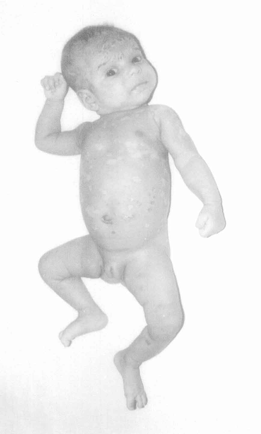
While it is uncommon for psoriasis to appear in neonatal period this undoubtedly does happen(l,2). There is strong association of early onset psoriasis with Class I and II HLA markers–including B13, Bw57, Cw6 and DR7. They are more likely to carry PSORS I gene. Since the initial lesions of psoriasis in neonatal period are most often in the diaper area, differentiation from other types of diaper eruption is difficult. Diaper dermatitis caused by the irritative effects of urine in the wet diaper area may imitate a psoriaform eruption. Psoriatic diaper rash is brighter red, better demarcated and often shinier than seborrheic dermatitis and lack the yellow scale that may be present with the latter. Two types of psoriatic rashes in the diaper distribution have been described namely "Localized psoriatic diaper rash" and "Psoriatic diaper rash with dissemination"(3). Both may be psoriasis or precursors to psoriasis in some infants. Our patient had features similar to the latter entity. As to the question of whether diaper psoriasis is an early manifestation of psoriasis, seborrheic dermatitis, or fungal infection, the possibility of psoriasis has been suggested because of a high incidence of family history of psoriasis, as was noted in our patient(3,4). There are reports of the development of true psoriasis many years later(5). The crucial factor in the etiology of diaper pustular psoriasis in our patient remains unclear. We believe this to be a case of psoriasis because of the skin biopsy findings and background of a family history.

Abhijeet Saha,
Prerna Batra,

Department of Pediatrics,
Mahatma Gandhi Institute of Medical Sciences,
Sewagram, Wardha 442 102,
Maharashtra, India.
E-mail: [email protected]

References

1. Lerner MR, Lerner AB. Congenital psoriasis: Report of three cases. Arch Dermatol 1972; 105: 598-601.

2. Watanabe M, Tabata W, Tagami H. Explosive diaper pustular psoriasis (Letter). Pediatr Dermatol 2002; 19: 564-565.

3. Morris A, Rogers M, Fischer G, Williams K. Childhood psoriasis: A clinical review of 1262 cases. Pediatr Dermatol 2001; 18:188-198.

4. Andersen SLC, Thomsen K. Psoriasis form napkin dermatitis. Br J Dermatol 1971; 84: 316-319.

5. Farber EM, Mullen RH, Jacobs AH, Nall L. Infantile psoriasis: A follow- up study. Pediatr Dermatol 1986; 3: 237-243.

Home

Past Issue

About IP

About IAP k

Feedback

Links

 Ahor Info.

  Subscription