|
|
Case Reports Indian Pediatrics 2006; 43:167-170 |
||||
|
Cutaneous Mucormycosis in Children |
||||
|
From the Amardeep Multispeciality Children’s Hospital & Research Center, Gujarat College Cricket Ground Road, Ellisbridge, Ahmedabad 380 006, Gujarat, India. Correspondence to: Dr. Amar Shah, Neonatal and
Pediatric Surgeon and Urologist, Amardeep Multispeciality Children’s
Hospital & Research Center, 65 Pritamnagar Society,
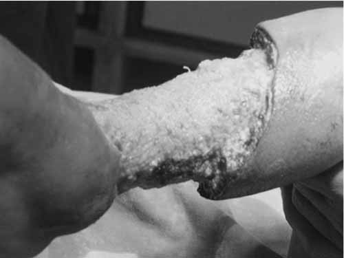
Mucormycosis is an uncommon infection caused by fungi of the order Mucorales, family Mucoraceae, class Zygomycetes(1). It was first described in humans by Paultaufi(2) in 1885. The common clinical forms include rhinocerebral, cutaneous, pulmonary, gastro-intestinal and disseminated. Cutaneous lesions may be either primary or secondary to dissemination from another site. Case Reports An eight-month-old child developed an abscess in the right leg following extravasation of an IV canula. Forty eight hours after the abscess was drained, he developed bluish purple discoloration proximal to the site of the incision. This gradually progressed following which the child was transferred to us for further management. On evaluation, he had extensive skin and subcutaneous tissue necrosis extending from the mid leg to the ankle. Distal pulsations were normal. Blood counts showed marked leucocytosis. Blood culture showed no growth. The child underwent excision of the necrotic skin and subcutaneous tissue. However skin erythema and subcutaneous necrosis continued to increase slowly and steadily with yellowish white cheesy material seen at the wound edges (Fig.1). Swabs from the wound grew Staphylococcus aureus sensitive to vancomycin. KOH stains showed mucormycosis. Histology showed subcutaneous necrosis with neutrophilic infiltrate and aseptate fungal hyphae. The child was started on single dose amphotericin B and six hourly vancomycin at 15 mg/kg/dose. Amphotericin B was started on day one with a test dose of 0.1 mg/kg/dose. This was further increased to 0.25 mg/kg/dose and then daily increased by 0.25 mg/kg to reach 1.5 mg/kg/dose. Amphotericin B was diluted in 5% dextrose in a way that the concentration does not exceed 0.1 mg/mL and infused over 4 hours. The infusion was protected from light. Vancomycin was diluted in Isolyte P and administered over a period of two hours. The child also underwent extensive debridement until the wound margins were visibly and histologically clear of mucor. Dressings were done with providone iodine. Amphotericin B was given for forty two days with weekly monitoring of complete blood count, serum potassium, creatinine and SGPT. Split skin thickness grafting was done following four weeks of treatment with amphotericin B. The child recovered well and discharged following 42-day regime of amphotericin B.
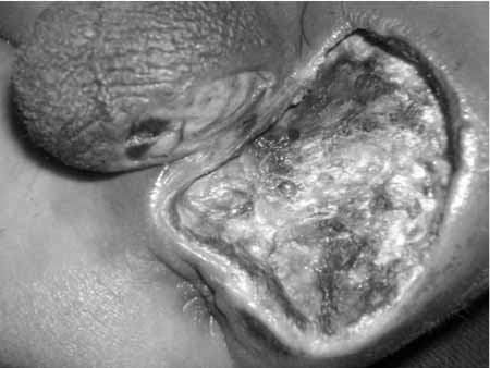
Case 2 A 2.0 kg boy was born at 34 weeks gestation by emergency cesarean section. The child was extensively resuscitated in the operating room for respiratory distress. He was commenced on corticosteroids and broad spectrum I.V. antibiotics. After 24 hours, a 1.5 cm diameter reddish black area was noted over the left perineum, 4 cm away from the anal verge. The area increased slowly and steadily to double the initial size in the next couple of days (Fig. 2) following which he was referred to us. The child underwent extensive debridement with excision of the necrotic skin patch. Debridement was not extended medially because of close proximity of the anal verge and the parents not consenting for a colostomy. Swabs from the wound and histology from the skin margins confirmed mucormycosis. Amphotericin B and IV. Vancomycin were commenced (dose and duration similar to those mentioned in case 1). Regular dressings were done with providone iodine. The wound eventually closed by secondary healing.
Discussion Mucormycosis is the most acute, fulminant and fatal of all fungal infections in humans. It presents most frequently in immuno-compromised hosts, but can occur in healthy patients in the presence of insignificant trauma(3). Predisposing factors like diabetes mellitus, neutropenia, corticosteroid therapy, malnutrition or burns make the host susceptible to this infection(4). Cutaneous inoculation from wooden tongue depressors(5) and cotton stockinettes(6) has also been reported. Not many cases of cutaneous mucormycosis in children are reported in the literature. In their review, Oh and Notrica found 18 cases of cutaneous mucormycosis in infants(7). Most of these patients were both premature and low birth weight. Corticosteroid therapy had been started on 12 patients because of respiratory distress. Many of them were also on broad spectrum antibiotics. The overall mortality rate in the series was 72%. Two different presentations of cutaneous mucormycosis have been described(8). The superficial type appears as vesicles or pustules that advance to ulceration and eschar formation usually in normal hosts. Rapidly progressing ulceration and dissemination in patients with decreased immunity is seen in gangrenous form. After inoculation, the fungus invades the vessels of skin and subcutaneous tissues causing necrosis. Clinically, this presents as small discolored area that becomes black and necrotic with surrounding erythema. Rapid dissemination to solid organs or blood may lead to fatal results. The first child was on broad spectrum intravenous antibiotics for pneumonitis. Skin damage following extravasation and drainage of the abscess created an environment conducive to the opportunistic organisms. The second child suffered respiratory distress after birth and was given corticosteroids to improve lung function. He was commenced on broad spectrum antibiotics. The skin damage could have been from trivial trauma encountered during resuscitation in the operating room. Neither of the two children were immuno-compromised or had any predisposing factors. Cutaneous mucormycosis is slowly growing and progresses in days as compared to the rapidly progressing Clostridium perfringens or Group A Streptococcal infections which progress in hours. Tissue biopsy with H&E staining is the main stay of diagnosis which shows characteristic broad, pauciseptate hyphae with branches occurring at right angles. Grocott methenamine-silver and periodic acid-Schiff stains may further improve visualization. Organisms from swab can be identified quickly with 10% KOH stain(9). Wound cultures are inadequate and should not be relied for a false sense of security(10). Blood cultures are rarely useful. Because of the infrequent and potentially fatal nature of the disease, a high index of suspicion and a low threshold for wound biopsy must be maintained. The recommended treatment has been systemic amphotericin (1-1.5 mg/kg/day) and extensive surgical debridement which may also lead to amputation in the worst scenarios(7,10). Aggressive treatment needs to be instituted without waiting for culture results as the end result depends upon the time period between recognition of necrotic lesion and institution of therapy. The debrided wound should be monitored for resolution of surrounding erythema and induration before definitive reconstruction. Addition of rifampin has shown to be helpful because of a synergistic activity with amphotericin(11). We started rifampin in the first case but found no significant advantage. Topical amphotericin B has also been shown to be helpful. However, availability and cost are a major restricting factor in its use in India. High index of suspicion, early diagnosis and rapid institution of therapy can improve the survival rate in children. The key to prevention appears to be appropriate skin care. However, the overall results in immuno-compromised patients remain poor. Contributors: AS was involved in the overall care and surgical management of the child and will act as a gurantor of the manuscript. SL and AS were involved in the surgical management. Funding: None. Competing interests: None.
| ||||
|
References | ||||
|
![]()