A 5-year-old girl presented with skin colored to whitish raised skin
lesions over the face, hands and trunk of three months duration. She
also had watering, pain and redness of the left eye. Her parents had
expired due to AIDS. On examination, she was asthenic with generalized
lymphadenopathy. Cutaneous examination revealed multiple, shiny, whitish
to skin colored, umbilicated papules (typical of molluscum contagiosum),
some of them were more than 2 cm in size. On puncturing, these lesions
expressed cheesy white material. They were distributed bilaterally
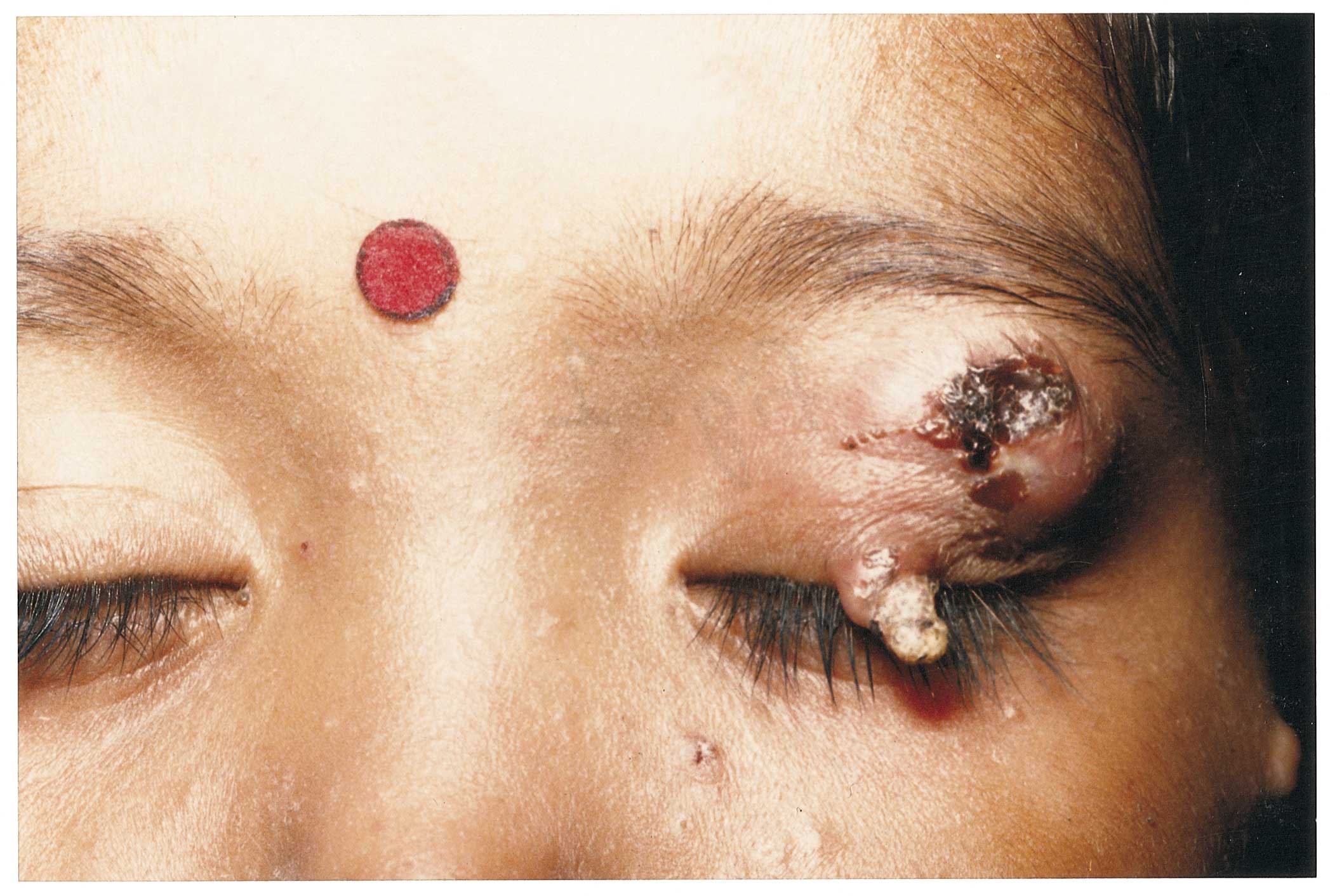
asymmetrically over the face, hands and trunk. A solitary firm horn like
projection, 1 cm in height was seen originating from the left upper
eyelid from a molluscum contagiosum lesion (Fig. 1). She was
diagnosed as having molluscum contagiosum and cutaneous horn. She also
had a positive serology for HIV. The skin lesions were treated with
extirpation and trichloroacetic acid cautery.
 |
|
Fig 1. Left eyelid margin showing a cutaneous
horn and adjoining skin having an infected molluscum contagiosum
lesion. |
Cutaneous horn (cornu- cutaneum) is the term coined
for horny skin excrescence, which in its form and consistency resembles
an animal horn in miniature. The paramount consideration while making a
clinical diagnosis is the height of the keratotic mass (at least one
half of its largest diameter). The important issue is not the horn
itself which is dead keratin, but rather the underlying condition, which
may be benign (seborrheic keratosis, viral warts, histiocytoma, inverted
follicular keratosis, verrucous epidermal nevus, molluscum contogiosum,
etc.), premalignant (solar keratosis, arsenical keratoses,
Bowen’s disease) or malignant (squamous cell carcinoma, rarely, basal
cell carcinoma, metastatic renal carcinoma, granular cell tumor,
sebaceous carcinoma or Kaposi’s sarcoma). Most commonly, they are single
and arise from a seborrheic keratoses lesion. They are encountered most
frequently on the face and scalp, but may occur on the hands, penis and
eyelids.
Devender Mohan Thappa,
Chandrashekhar Laxmisha,
Department of Dermatology and STD,|
Jawaharlal Institute of Postgraduate Medical
Education and Research (JIPMER),
Pondicherry 605 006, India.
E-mail: dmthappa@satyam.net.in
