Paraesophageal hernia consists of displacement of the
stomach into the thoracic cavity alongside the esophagus, which remains
in its normal position. This is an anatomical defect of the hiatus
without any derangement of the gastroesophageal sphincter, which clearly
distinguishes it from other types of hiatal hernia. Anderson(1) reported
paraesophageal hernia to be rare in children, particularly in the
neonatal period. The problems of gastro-esophageal reflux and sliding
hernia in pediatric age group over-shadow the pathology of the
esophageal hiatus of the diaphragm in this age group. Its presentation
in neonatal period can be confused with the possibility of esophageal
atresia(2), or esophageal web. The contrast study for esophagus and
stomach is diagnostic in this disease, but careful interpretation of
plain X-ray of chest can also raise the suspicion of the disease.
Two neonates with paraesophageal hernia are reported, one with the
mesenterico-axial volvulus and herniation of spleen along with the
stomach, and second neonate without volvulus.
Case Reports
Case 1
A one-day-old male neonate presented with minimal
respiratory distress since birth. Physical examination was essentially
normal except for mild tachypnea (50/min). Infantogram revealed presence
of gaseous shadow in the left paravertebral region of lower chest, as
well as in the subdiaphragmatic area, with presence of nasogastric tube
in the subdiaphragmatic stomach (Fig. 1). An air contrast study
for esophagus and stomach showed the presence of stomach in the lower
chest on lateral infantogram. Exploratory laparotomy through left
subcostal incision revealed a paraesophageal hernia.
 |
 |
Fig 1. Infantogram (A-P view) of
case 1 showing a gas filled cystic lesion (arrow) in the left
lower chest.
|
Fig 2. Infantogram of case 2
with catheter in lower esophagus at T7 vertebra level (small
arrow) and intrathoracic stomach in the right lower chest (large
arrow). |
There was no volvulus of stomach, and stomach could
be reduced into the peritoneal cavity with ease. The esophageal hiatus
was narrowed using interrupted Vicryl sutures, and gastropexy was added
to the procedure. This baby made an uneventful recovery and is now three
years old and asymptomatic.
Case 2
A seven-day-old neonate weighing 3 kg was referred to
us with a suspected diagnosis of esophageal atresia due to excessive
salivation, regurgitation of feeds and 2 episodes of cyanotic spells.
Physical examination did not reveal any overt signs, but an orogastric
tube was arrested at 15 cm from the gum margin. Plain X-ray of
the chest with orogastric tube in situ demonstrated the presence
of tip of the tube at seventh thoracic vertebral level as well as the
presence of an abnormal gas shadow in the right para vertebral region in
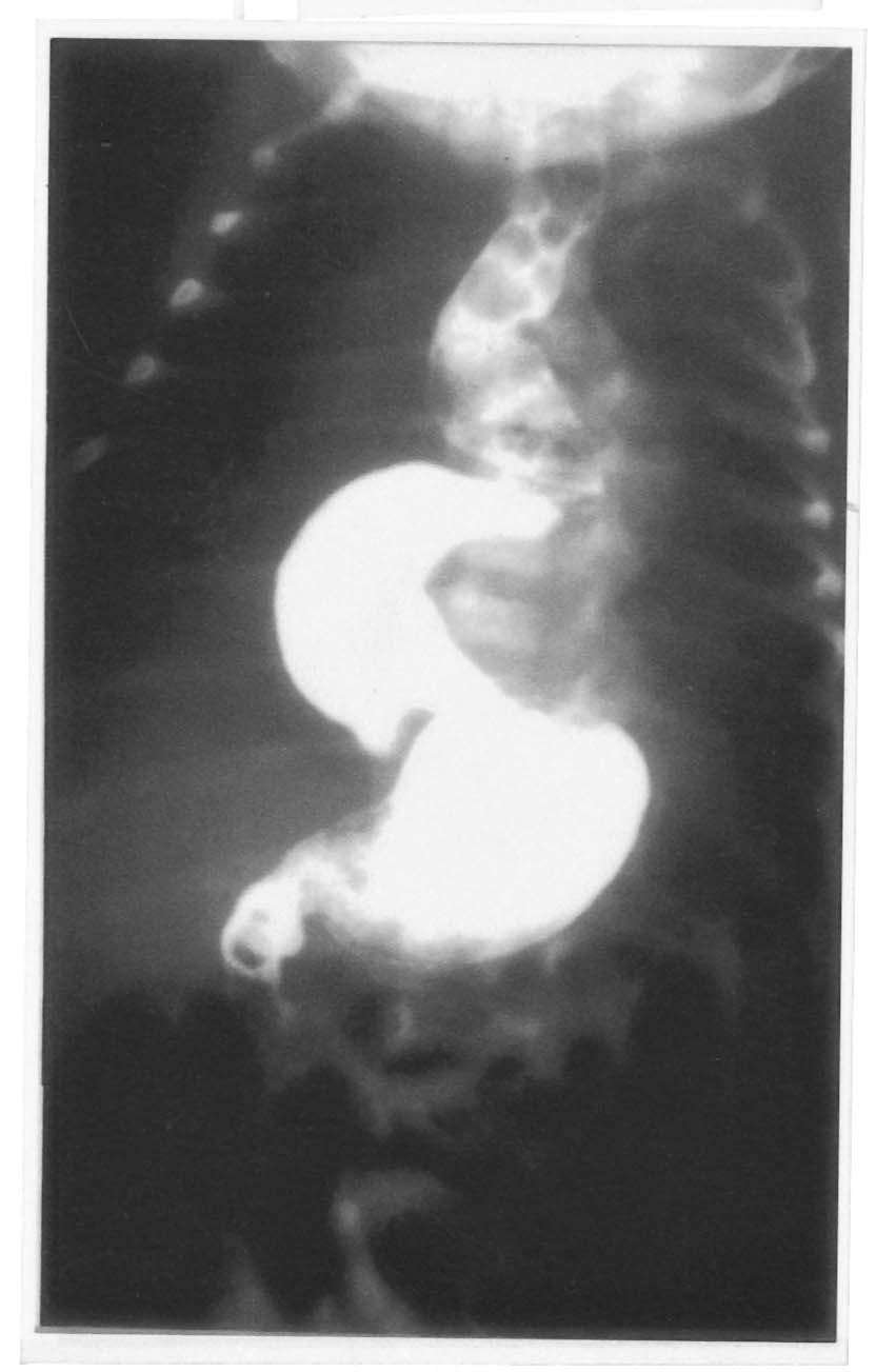
the lower chest (Fig. 2). A contrast study using water-soluble
contrast revealed dilatation of esophagus and an intrathoracic stomach (Fig.
3). The stomach was in a state of volvulus and a small quantity of
contrast passed distally into the duodenum. A laparotomy was performed;
surprisingly the spleen was also herniated alongside the stomach. The
herniated contents were reduced into the peritoneal cavity with
derotation of the volvulus. The esophageal hiatus was about 3 cm in
size, which was narrowed with interrupted sutures and a fundopexy
per-formed with fixation of the gastric fundus to the left hemidiaphragm.
An uneventful post-operative recovery followed and the child is doing
well at one-year follow-up.
 |
|
Fig. 3. Upper GI contrast study-showing
mesenterico-axial volvulus of stomach: A small quantity of
contrast passing distally into duodenum. |
Discussion
Paraesophageal hernia is one of the several known
defects of the diaphragm. Paraeso-phageal hernias are seen at all ages
throughout life, but rarely present in the neonatal period. However,
paraesophageal hernia has also been reported in siblings(3). A variety
of symptoms may occur, but it is common that the symptom complex is
indicative of upper alimentary tract obstruction, with or without a
cyanotic episode. In patients with the associated gastric volvulus, the
symptomatology may indicate an esophageal atresia(2), as was suspected
in case 2 of our study. In these neonates an orogastric tube gets
arrested at 14 or 15 cm from the gum margin (Fig. 2), a level
lower than that usually seen in esophageal atresia. An upper
gastrointestinal tract contrast study usually shows the presence of
stomach in the lower thorax. Rarely, an infant with neonatal Marfan
syndrome can present with hiatus/para esophageal hernia with or without
gastro-esophageal reflux(4).
A plain X-ray of chest in these neonates needs
to be viewed carefully as the gastric shadow can be seen in lower chest,
by the side of esophagus and separate from the pulmonary shadow (Figs.
l and 2). In both the neonates a contrast study was performed for
delineation of anatomy of esophagus and stomach, and a correct
preoperative diagnosis could be made.
Case 2 was unique in that the spleen was herniated
alongside the stomach into the thoracic cavity, to our best of knowledge
this has not been reported earlier. Both the neonates had isolated
paraesophageal hernia and are doing well 1 year and 8 months
postoperatively. The purpose of this paper is to draw attention to the
possibility of paraesophageal hernia in neonatal period and to suspect
this disease entity even on plain x-ray of chest.
Contributors: DK and RS designed the study. KLNR
helped in the preparation of the manuscript.
Funding: None.
Competing interests: None stated.
