|
|
|
Indian Pediatr 2018;55: 1087-1088 |
 |
Coexisting Congenital
Subglosso-palatal Membrane and Tongue Dermoid in a Neonate
|
|
Preeti Tiwari 1,
Vaibhav Pandey2
and Jayanto Tapadar3
From Departments of 1Oral and
Maxillofacial Surgery, and 2Paediatric Surgery,
Institute of Medical Sciences – Banaras Hindu University, and 3Samayan
Hospital; Varanasi, Uttar Pradesh, India.
Correspondence to: Dr Preeti Tiwari, Department of
Oral and Maxillofacial Surgery, IMS – BHU, Varanasi 221 005, UP, India.
Email:
[email protected]
Received: February 04, 2018;
Initial review: May 19, 2018;
Accepted: October 12, 2018.
|
Background: Neonatal respiratory distress due to coexisting
subglosso-palatal membrane and tongue dermoid has not been reported yet.
Case characteristics: A newborn with respiratory distress having
a membrane in the oral cavity. Excision of membrane revealed a tongue
mass with cleft palate, obstructing the nasopharynx completely. Elective
ventilation was followed by excision of mass. Outcome: The child
was cured with uneventful course at follow-up of six months. Message:
Co-existing congenital anomalies causing airway obstruction may be
missed in presence of subglosso-palatal membrane.
Keywords: Infant, Respiratory distress, Stridor.
|
|
C
ongenital anomalies affecting the oral cavity are
rare. Often these can cause respiratory distress, aspiration, and
bleeding [1]. It is unusual to have various congenital pathologies of
the oral cavity in same patient, resulting in complexity in presentation
and management. A congenital mass in the oral cavity can be a cause of
respiratory distress [1]. Similarly, the subglosso-palatal membrane
which is thought to be a remnant of the buccopharyngeal membrane is also
known to be associated with respiratory distress [2]. We share our
experience and challenges faced during management of a neonate with
severe respiratory distress, who had a co-existence of these two birth
defects.
Case Report
A full-term male baby weighing 2.8 kg was delivered
by spontaneous vaginal delivery to an unbooked primigravida mother at a
peripheral center. The child presented to us with severe respiratory
distress with a heart rate of 180/min, a respiratory rate of 60/min
along with inspiratory stridor and subcostal retraction. The air entry
was diminished but equal on both the sides. The oxygen saturation on
high flow oxygen, via high flow nasal cannula, at the rate of
10L/min, was 90-95%. On oral examination, there was a membrane extending
from the floor of the mouth to the junction of the soft and hard palates
with its lateral extension up to the molar trigone bilaterally. Nasal
suction was performed to clear the secretions and to rule out associated
choanal atresia.
The child deteriorated rapidly and progressed to
respiratory failure. The respiratory rate fell to 20-30/min and
saturation dropped to 75-80% on high flow oxygen. A diagnosis of the
sub-glossopalatal membrane with severe respiratory distress and
respiratory failure due to upper airway obstruction was made. The bag
and mask ventilation was started. The distended stomach was decompressed
with a infant feeding tube, though there was difficulty in negotiating
it through the nasopharynx.
As the bag and mask ventilation was ineffective, an
emergency excision of the membrane was performed. On excision of the
membrane, a mass was seen to be arising from the dorsum of the tongue.
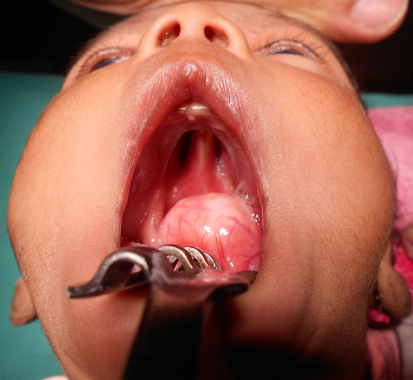
The lesion was nearly filling the oropharynx and the nasopharynx (Fig.
1). The tongue was gently pulled out of the mouth, and the child was
intubated to secure the airway. The child was shifted on the ventilator.
After 24 hours, the baby improved and was extubated after which he was
nursed in prone position.
 |
|
Fig. 1 The mass arising from the
dorsum of tongue with palatal cleft.
|
A computed tomography scan showed a cystic mass
arising from the dorsum of the tongue extending into oropharynx and
nasopharynx, and associated with cleft palate. Echocardiography and
abdominal ultrasound were normal. An elective excision of the mass was
planned. Per-operative, the mass was tense, cystic and lobulated with
extension into the substance of tongue. The upper portion of the mass
was filling the space of palatal cleft while the lower and posterior
portions were occupying the nasopharynx and oropharynx. Primary closure
of dorsum of the tongue was achieved after the complete excision of the
mass. The postoperative course was uneventful. The child is awaiting
cleft palate repair. Histopathology of the mass showed features
suggestive of a dermoid cyst.
Discussion
Congenital anomalies of the oral cavity are rare. As
these result from abnormal development at a very early stage, they can
be associated with multiple complications. Further, it is rare to find
multiple congenital lesions affecting the oral cavity in one patient. A
congenital lesion in the oral cavity may cause upper airway obstruction
and lead to respiratory distress at the time of birth [2]. Similarly,
the sub-glossopalatal membrane can compromise the upper airway patency
and present with respiratory distress [3]. Both lesions presenting
simultaneously in one patient is extremely unusual.
In this case, the child had respiratory distress due
to upper airway obstruction caused by congenital anomalies. Upper airway
in neonates may be blocked due to anomalies like choanal atresia, tumors
such as glioma, encephalocele, teratoma or dermoid, vocal cord
paralysis, and subglottic stenosis [1,5]. Many of these cases may
require emergency surgical intervention for restoration of a secure
airway [1]. Subglossopalatal membrane is a remnant of the
buccopharyngeal membrane [3]. As neonates are obligate nasal breathers,
the subglosso-palatal membrane may not cause respiratory distress in all
cases [5]. This created a diagnostic dilemma in our case, and we looked
for associated choanal atresia, as this association has previously been
reported [6].
Congenital tongue mass can cause upper airway
obstruction and child may have respiratory distress at birth [2]. The
development of tongue is one of the earliest events in fetal life
(4th–5th week) and can affect the development of palate and other
maxillofacial structures [7]. Thus, a mass on the dorsum of the tongue
can protrude into the defect between the palatal shelves (palate
develops between 6th and 7th week) and prevent their fusion, and
therefore, can cause palatal cleft [8]. Further, as the musculature of
the tongue is formed there can be fusion, entrapment, and proliferation
of epithelial debris, which can lead to the development of lingual
dermoid cyst [9]. This can explain the infiltration of dermoid into the
substance of tongue in our case [10]. The development of tongue and
glossopharyngeal membrane and other maxillofacial structure overlap
closely with each other. Thus, one anomaly may lead to the development
of others.
Through this case, we documenting rare co-existence
of subglosso-palatal membrane, cleft palate and dorsal tongue dermoid.
Subglosso-palatal membrane excision should be performed in an emergency
if the child is not improving on high flow oxygen because some other
associated anomaly can be missed due to blocked view.
Contributors: All authors contributed to case
management and manuscript.
Funding: None; Competing interest: None
stated.
References
1. Grossfeld JL, Ballantine TVN. Surgical respiratory
distress in infancy and childhood. Curr Probl Pediatr. 1976;6:1-64.
2. Celik M, Akkaya H, Arda IS, Hiçsönmez A.
Congenital teratoma of the tongue: a case report and review of the
literature. J Pediatr Surg. 2006;41:e25-8.
3. Nakajima T, Takahashi MTS. Subglosso-palatal
membrane. Plast Reconstr Surg. 1979;63:574-6.
4. Kumar A, Bhatnagar V. Respiratory distress in
neonates. Indian J Pediatr. 2005;72:425-8.
5. Coates H. Nasal obstruction in the neonate and
infant. Clin Pediatr. 1992;31:25-9.
6. Pandey V, Tiwari P, Tapadar J, Gangopadhyay A.
Subglosso-palatal membrane a rare cause of neonatal respiratory
distress: A case report. J Indian Assoc Pediatr Surg. 2014;19:109-11.
7. Hong SJ, Cha BG, Kim YS, Lee SK, Chi JG. Tongue
growth during prenatal development in Korean fetuses and embryos. J
Pathol Transl Med. 2015;49:497-10.
8. Poretti A, Vitiello G, Hennekam RCM, Arrigoni F,
Bertini E, Borgatti R, et al. Delineation and diagnostic criteria
of oral-facial-digital syndrome type VI. Orphanet J Rare Dis. 2012;7:4.
9. Raewyn C, Paul W. Management of congenital lingual
dermoid cysts. Int J Pediatr Otorhinolaryngol. 2010;74: 567-71.
10. Turki IM, Mouaffak-Zidi Y, Rajhi H, Triki H, Naija S, Ben JS,
et al. A case of large dermoid cyst of the tongue. Egypt J Ear Nose
Throat Allied Sci. 2011;12:171-4.
|
|
|
 |
|
