A 14-year-old girl, resident of Uttar Pradesh, India, presented to us
with multiple lesions appearing in crops recurrently for past 1 year.
The lesions appeared over the normal appearing skin with a frequency of
6-7 reddish raised lesions per day and healed spontaneously in a period
of 10-12 days leaving behind hyperpigmentation. Each episode was
associated with high fever and generalized malaise. In this episode,
patient noticed pustulation followed by ulceration over the lesion
within 5 days of appearance of the crop. There was history of recurrent
epistaxis for past one year and ear discharge prior to current
exacerbation of lesions. There was no history suggestive of motor
weakness, pain or edema over limbs, redness of eye, photophobia, frothy
urine, palpitation, or dyspnea on exertion. Lesions of various
morphology in form of pustules, nodules and plaques with ulceration and
erosions in various stages of healing were present in a generalized
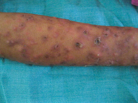
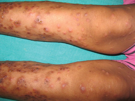
fashion all over the body, predominantly involving the limbs (Fig.
1) and trunk. Diffuse infiltration was present over face and
ears. Multiple peripheral nerves were thickened bilaterally. However,
there was no glove and stocking anesthesia. The patient had received
multiple courses of antibiotics with a diagnosis of recurrent
furunculosis. Hematological and biochemical investigations were within
normal limits and histopathology from nodular lesion was suggestive of
erythema nodosum leprosum necroticans. Multibacillary multidrug therapy
was initiated along with oral prednisolone 40 mg daily.
(a) |
(b) |
|
Fig. 1 Multiple erythematous nodular
lesions along with pustules and ulcers with hemorrhagic crusting
over forearms (a), and over legs (b).
|
| |
The differential diagnoses for ulcerated nodules with
fever would include furunculosis, mycobacterial infections and childhood
vasculitis like childhood polyarteritis nodosa, benign cutaneous
polyarteritis nodosa and wegeners vasculitis. Cutaneous polyarteritis
nodosa is a relapsing chronic disease that presents with crops of
painful, erythematous, subcutaneous nodules predominantly over the lower
legs, with associated urticaria, livedo reticularis, peripheral
gangrene, myalgia, arthralgia, non-erosive arthritis and peripheral
neuropathy. Therefore it may closely mimic erythema nodosum leprosum and
careful examination will detect diffuse infiltration especially over the
face and nerve thickening or deformity suggestive of lepromatous
leprosy. This case highlights the need to retain the focus on leprosy so
that physicians can ensure early detection and treatment, including
screening for family contacts, to reduce morbidity and decrease
community burden of leprosy.


