|
|
|
Indian Pediatr 2016;53: 1133 |
 |
Mongolian Spots in GM1 Gangliosidosis
|
|
Naveen Kumar Bhardwaj and *Daisy Khera
Department of Pediatrics, AIIMS, Jodhpur, Rajasthan,
India.
Email: [email protected]
|
|
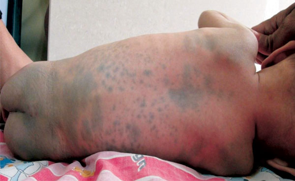
A 6-month-old girl born out of second degree
consanguinity presented to us with multiple greyish-blue, well- to
ill-defined macules and patches of varying sizes, involving chest,
abdomen, back and buttocks (Fig. 1). Child had global
developmental delay with developmental age of 2-3 months. Physical
examination revealed coarse facial features, hepatomegaly and
pansystolic murmur. Further investigations revealed normal fundus,
hypertrophic cardiomyopathy and tiny persistent ductus arteriosus.
a-Galactosidase
activity in peripheral blood leukocytes was significantly reduced,
indicative of type I (infantile) GM1 gangliosidosis.
 |
|
Fig. 1 Multiple greyish-blue macules
and patches of varying sizes involving whole back and buttocks.
|
Typical and limited mongolian spots are benign blue
or slate gray macular lesions of varying sizes, most commonly located on
lumbosacral region. Mongolian spots usually fade during the first few
years of life, but they occasionally persist. Those associated with
inborn errors of metabolism are extensive and show no sign of
resolution. Extensive Mongolian spots are most frequently associated
with GM1 gangliosidosis, Hurler-Scheie syndrome, Niemann-Pick disease,
Hunter syndrome and a-mannosidosis.
The findings of generalized mongolian spots in an infant may represent
underlying storage disorders thereby allowing identification of families
at risk.
|
|
|
 |
|
