|
|
|
Indian Pediatr 2013;50: 1180 |
 |
Grouped Vesicular Lesions in an Infant
|
|
Avijit Mondal, Neloy Sinha and *Piyush Kumar
Department of Dermatology, College of Medicine and JNM
Hospital;
and *Katihar Medical College and Hospital Karim Bagh,
Katihar - 854105,
Bihar, India.
Email: [email protected]
|
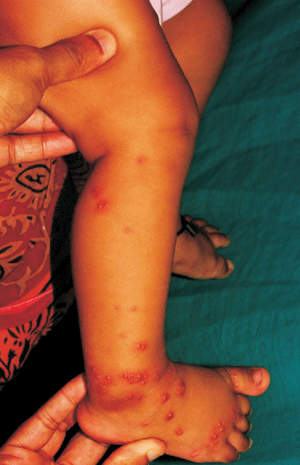
An 8-month-old female infant presented with vesicular
lesions on the right lower extremity for 2 days. There were
grouped vesicles on an erythematous base over right leg,
dorsum of right foot and sole, distributed in the L5 and S1
dermatomes (Fig 1). The baby was irritable, but
afebrile. The baby was born at term and by normal vaginal
delivery and her birth weight was 2.5 kg. Postnatal period
was uneventful. Her developmental milestones were within
normal range. There was history of maternal varicella
infection during 3rd months of pregnancy. Tzanck smear from
the lesions showed mononuclear and multinucleated
acantholytic cells with ground glass nuclei, consistent with
Tzanck cells. Based on history and clinical findings,
diagnosis of Herpes zoster was made. The infant was treated
symptomatically with topical calamine lotion and oral
antipyretic. The lesions crusted in 1 week and resolved
completely in 2 weeks, without any sequelae.
 |
|
Fig 1
Grouped vesicles on an erythematous base, present
unilaterally on right lower extremity. Note central
umbilication in some of the vesicles.
|
Herpes Zoster (HZ) results from
reactivation of varicella Zoster virus (VZV) that entered
the cutaneous nerves during an earlier episode of
chickenpox, traveled to the dorsal root ganglia, and
remained in a latent form. Age, immunosuppressive drugs,
lymphoma, fatigue, emotional upsets, and radiation therapy
have been implicated in reactivation of the virus, which
subsequently travels back down the sensory nerve, infecting
the skin. Reactivation of latent VZV infection is very rare
in childhood, more so in infants. Infantile HZ is more
commonly associated with intrauterine VZV infection than
postnatal infection. HZ in children is considered common in
immunocompromised babies, but can occur in immunocompetent
children as well. The diagnosis can usually be made on
clinical grounds. Tzanck smear may support the clinical
diagnosis. The differential diagnoses are zosteriform herpes
simplex infection (no radiating pain, small vesicles of
almost uniform size, less number of groups of vesicles, and
more likely to recur) and contact dermatitis (history of
contact, and presence of papules, pustules, scaling and/or
epidermal necrosis). HZ is a self-limited cutaneous eruption
in children and treatment usually consists of supportive
care with antihistamines and antipruritic agents. Systemic
antiviral drug is usually recommended in severe disseminated
VZV infection, ophthalmic VZV infection and in
immunocompromised patients.
|
|
|
 |
|
