A 4-year-old girl born of non-consanguinous marriage,
presented with generalized vesiculobullous eruptions for a
duration of 5 months. To start with, few small fluid filled
lesions appeared over abdomen, then gradually involved the
rest of the trunk, upper and lower extremities and also
face, palms and soles. The lesions gradually increased in
size and ruptured over few areas forming erosions. Patient
also had burning sensation of mouth during food intake.
There was no history of trauma-induced blisters. There was
no history of drug intake prior to the onset of blisters and
no history of photosensitivity was obtained. There was no
past history or family history of similar illness or any
other significant medical or surgical history.
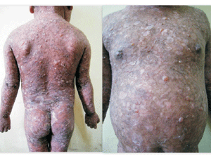
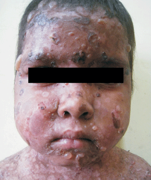
Cutaneous examination revealed multiple
tense vesicles, bullae and crusted erosions all over the
body including face, palms and soles. (Fig. 1
and 2) Nikolsky’s sign and bulla spread sign were
negative. Older lesions healed with hypopigmentation without
increasing in size at the periphery. Mucous membrane
examination revealed erosions over hard palate and buccal
mucosa. There was no regional lymphadenopathy. Routine
laboratory tests were normal. Tzanck smear was negative.
 |
 |
|
Fig. 1 Numerous blisters
and erosions over the body.
|
Fig. 2 Blisters and
erosions over a cushingoid facies.
|
Histopathological examination revealed
subepidermal blister with neutrophils and fibrin. Dermis
showed mild inflammatory infiltrate composed of polymorphs
and few eosinophils. Provisional diagnosis of chronic
bullous disease of childhood (CBDC) was made. Direct
immunofluorescence (DIF) showed linear deposition of IgG and
C3 at the dermoepidermal junction. DIF confirmed the
diagnosis of bullous pemphigoid, childhood variety.
Close clinical differential diagnoses
include CBDC, Epidermolysis bullosa, porphyria. String of
pearls configuration of bullae which is seen in CBDC was not
present in our case. There was no history of trauma induced
blister as well as family history and nail dystrophy was
absent. Thus, Epidermolysis bullosa was excluded. Porphyria
was excluded by absence of photosensitivity, urine
discoloration, systemic features or scarring. Finally DIF
confirmed our diagnosis of bullous pemphigoid.
Bullous pemphigoid (BP) is the most
common autoimmune subepidermal blistering disease. The
disease typically presents in the elderly, with an onset
after 60 years of age. Childhood variant of bullous
pemphigoid is very rare.
Systemic corticosteroids represent the
first line treatment at a dose of 0.5–1 mg/kg/day. Other
immunosuppressive drugs frequently used are azathioprine,
methotrexate, cyclophosphamide, cyclosporine and
mycophenolate mofetil. The combination of nicotinamide and
tetracycline has been found to be useful. Our patient did
not respond to sulphones but responded well to prednisolone
which also reinforced our diagnosis.

