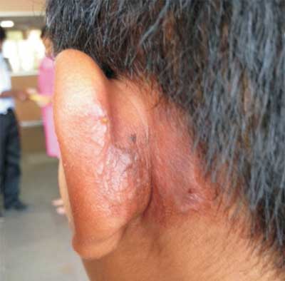
An eleven- year -old boy presented with erythematous lesion
over the left post auricular region for one day. There was
history of an insect seen over the area of rash on previous
night. On examination, he was afebrile and had erythematous
rash with vesicles over the posterior aspect of the left
auricle and mastoid area (Fig. 1). The findings of
systemic examinations were normal. His complete blood count
was within normal limits. Based on the history, presence of
typical kissing lesions a diagnosis of paederus dermatitis
was made. The child was treated with topical steroid
ointment. Skin lesions healed completely within one week.
 |
|
Fig.1 Vesicles over
auricle.
|
Paederus beetles have been associated
with outbreak of dermatitis in various countries. Adult of
these beetles are usually 7-10 mm long and 0.5 mm wide. They
have black head and red thorax. Dermatitis is caused by
paederine which is released on crushing the insect on the
skin. The rash appears 24 hours after contact. A striking
feature is the presence of kissing lesions that occur
wherever apposition of skin is possible (e.g. flexure
of the elbow, adjacent surfaces of the thigh). Clinical
appearance of paederus dermatitis may be confused with
herpes zoster, acute allergic contact dermatitis, liquid
burns, millipede dermatitis and phytophoto dermatitis. There
are many similarities between paederus dermatitis and
phytophoto dermatitis including linear asymmetric areas of
erthyema, possible blister formation and dyspigmentation.
With phytophoto dermatitis, there is a history of exposure
to light sensitizing biological substance such as lime or
fig. The characteristic linear appearance of the lesion, the
presence of kissing lesions and their predilection for
exposed areas differentiate paederus dermatitis from other
above mentioned conditions. Histopathology may support the
diagnosis of paederus dermatitis. The cases should be
managed with initial washing the area with soap and water
followed by topical steroid ointment.


