|
|
Images in Clinical Practice Indian Pediatrics 2006; 43:1103-1104 |
||||
|
Incontinentia Pigmenti |
||||
|
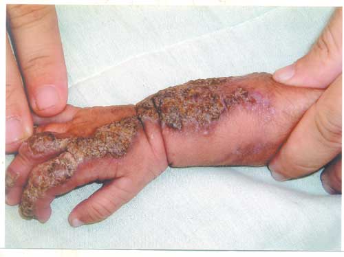
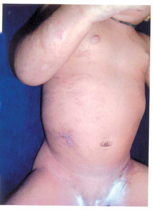
Incontinetia pigmenti (IP) (syn: Bloch-Sulzberger disease) is a rare hyperpigmentary disorder with a X-linked dominant inheritance. It is characterized by four phases-vesicular, verucous, pigmentary and hypopigmentary stage. In 80% cases other defects are seen in the form of dental, skeletal, nail anomalies, microcephaly, seizures, psychomotor retardation, strabismus, opitc nerve atrophy, retinal detachment, cataracts etc. The skin lesions are benign and follow lines of Blaschko. These lesions resolve by the age of one year but pigmentary stage may last for years. Pigmentation fades away by adulthood, leaving no sequelae. Blood levels of IgE are raised with eosinophilia.
The blistering stage of the lesion needs to be differentiated from Herpes simplex and Bullous Impetigo. The lesions are linear and in clusters in classical Incontenentia Pigmenti Warty phase needs to be differentiated from linear epidermal birthmarks or warts. Hyperpigmentary stage needs to be differentiated from moles and other causes of hyperpigmentation. Hyperpigmentation of Incontenentia Pigmenti is classically in whorls. Skin lesions of Incontinentia pigmenti are self limiting. Treatment is usually symptomatic in the form of emollients and topical antibiotics if required for infection. Shailaja Mane, |
![]()