Papillon-Lefevre syndrome (PLS) is a rare autosomal
recessive disorder of keratinization. Its reported incidence is 1-4 per
million and both the sexes are equally affected(1). It is characterized
by palmo-plantar hyperkeratosis, periodontopathy and premature loss of
deciduous as well as permanent dentition. It manifests between 1-5 year
of life and the patient becomes edentulous in the early teens. Another
component of PLS is asymptomatic ectopic calcification in choroid plexus
and tentorium. Although this has been taken as a cardinal feature, but
being inconsistent it is not considered important for the diagnosis.
About 20% of these patients also show an increased susceptibility to
infections due to some dys-function of lymphocytes and leukocytes(2).
The diagnosis is mainly clinical.
We describe here two cases of PLS with classic
clinical features.
Case 1
This 15-year-old girl presented with palmoplantar
hyperkerotosis since the age of four years and total loss of teeth by
the age of 12 years. She was the first child born to apparently healthy
non-consanguineous parents after an uneventful pregnancy and birth. Her
five younger siblings were reportedly normal. History revealed that her
deciduous teeth had erupted normally but exfoliated gradually by the age
of 4-5 years. Similarly, her permanent teeth too were lost prematurely
after erupting normally. There was history of recurrent swelling of gums
and foul breath followed by loosening and exfoliation of teeth. At the
age of four years, her parents also noticed a progressive thickening of
palmoplantar skin. It was associated with marked aggravation of erythema,
scaling and dryness during the eruption of teeth, and had improved after
complete exfoliation of dentition.
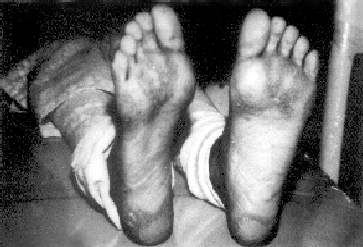
On examination, there was diffuse palmoplantar
keratoderma, transgradiens extending up to dorso-lateral aspects and
tendo-achillis area (Fig. 1). It was diffuse and severe on soles
while punctate and striate on palms. No other cutaneous lesion or
abnormality of hair, nails or sweating was seen. Intraoral examination
revealed com-pletely edentulous ridges with normal overlying mucosa. The
two mobile mandibular second molars showed surroun-ding inflammation and
gingival recession. Her systemic examination and routine laboratory
investigation including chest and skull X-ray films were normal.
Alveolar healing occurred normally after the extraction of mobile
mandibular molars. She was provided artificial dentures subsequently.
 |
|
Fig. 1. Plantar keratoderma with
transgradiens |
Case2
This 10-year-old girl was referred from the dental
clinic for the evaluation of cutaneous lesions. She was the second child
born normally to non-consanguineous parents. There was no family history
of similar complaints. History revealed early loss of her deciduous
teeth after normal eruption and development of palmoplantar
hyperkeratosis at the age of two years.
Cutaneous examination revealed palmo-plantar
keratoderma, more on pressure areas with transgradiens extending up to
dorsolateral aspects and tendoachillis area. Sweating, hair and nails
were normal. Intraoral examination revealed painful, swollen, bleeding
gums and fetid odor. The involved gingiva was bright red and margins
were hyperplastic. There was loss of gingival stippling and bleeding
occurred on probing the involved gums. She was wearing an acrylic splint
for multiple loose teeth. The following loose permanent teeth (FDI
notation) were present: 15, 14, 12, 11, 21, 22, 24, 25, 26, 36, 35, 34,
33, 32, 31, 41, 42, 43, 44, 45. On removal of acrylic splint for oral
prophylaxis, the mandibular central incisors exfoliated spontaneously.
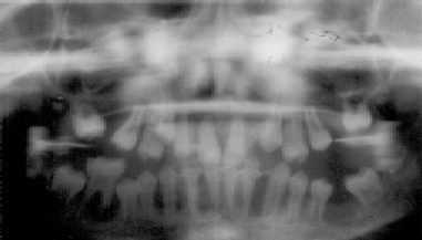
The orthopantograph showed unerupted all second
molars and maxillary canines. There was rapid loss of alveolar
attachment and the affected teeth lacked osseous support. The alveolar
bone around the mobile teeth was devoid of definable lamina dura and
showed indurated periodontal membrane pockets. An extensive alveolar
bone loss was noted, giving the teeth a "floating-in-air" appearance (Fig.
2). Other systemic exami-nation, routine laboratory investigations,
X-ray films for skull and chest were normal. The acute
inflammation improved to some extent with peridontal cleaning and
antibiotic therapy. However, the patient did not turn up for further
follow up.
 |
|
Fig. 2. Floating-in-air appearance of
teeth on orthopantograph. |
Discussion
It is generally accepted that PLS is a manifestation
of homozygosity of autosomal recessive genes with consanguinity as a
contributive factor. A major gene locus for PLS has been mapped to a 2.8
cm interval on chromosome 11 q 14 and inheritance of mutations of the
cathepsin C gene is found in homozygotes of PLS(3). Variable clinical
features and parental non-consanguinity suggest some heterogeneity and
variable expressibility of the condition. Both our patients were
non-consanguineous and appear to have inherited the condition
dominantly.
Swelling of gums and severe periodontitis becomes
evident in first year of life itself and loss of primary teeth occurs by
the age of 3-4 years. The periodontal inflammation subsides after
exfoliation of the deciduous teeth. All the erupted permanent teeth are
then lost after periodontal inflammation by the age of 13-16 years.
Later the third molars also undergo the same fate. Severe resorption of
alveolar bone gives the teeth a " floating-in-air" appearance on dental
X-ray film. Our both patients showed these classic events of
gingivitis, periodontotitis and precocious loss of deciduous as well as
permanent dentition at the age of 15 and 10 years. The gingival
inflammation subsided and alveolar healing occurred after the extraction
of second molars in Case l.
Palmoplantar keratoderma, severe on soles, usually
manifests as erythemato-keratotic and sharply demarcated lesion with a
tendency for worsening in winter. It may be punctate, striate or diffuse
with trans gradient. Hyperkeratotic lesions may also be seen on knees,
elbows and achillis tendon areas. Maximum severity of hyperkeratosis
coincides with severity of periodontal disease. It tends to improve by
puberty and after exfoliation of all the permanent teeth(4). Both are
probably interrelated, as the patients are edentulous by that time. Our
patients had typical palmoplantar keratoderma but only the first patient
showed its severity coinciding with gingival inflammation and tendency
towards improvement after exfoliation of all the teeth.
Hair are usually normal and nails may show
onychodystrophy and transverse grooving. Claw like phalanges with convex
nails (arachnodactyly) and osteolysis(5) described in PLS, are perhaps
its variants. No hair and nail abnormalities were observed in our
patients.
It is usually not necessary to treat cutaneous
lesions unless they interfere with patient’s activities. Frequent
periodontal cleaning, oral hygiene instructions and antibiotic therapy
only delay the shedding of teeth. Early extraction of teeth too has been
advocated to prevent bony loss. Moreover, this allows solid base for
subsequent use of artificial dentures. Etretinate, isotretinoin and
acitretin have all been successful in improving the cutaneous as well as
gingival lesions. However, normal dentition is observed with retinoids
only when given before the onset of permanent teeth at 5 years of
age(6).
Gingival infection, abscess formation, loss of
alveolar bone and destruction of the periodontal ligament are probably
the causative factors in shedding of teeth. A multitude of etiologic
factors appear to be involved in PLS. Page and Baab(7) opined that the
defect seems to be with the periodontal component especially the
cementum and as the teeth lose their last close relation to bone, the
intensity of inflammation subsides. According to Preus(8) the hereditary
defect perhaps lie at the epithelial surface barrier leading to reduced
defense against virulent periodontopathogens. Furthermore, the severity
of palmoplantar hyperkeratosis coinciding with that of periodontal
disease and successful retention of teeth by elimination of potential
pathogens is also suggestive of their association. There may also exist
a direct relation between PLS and psoriasis; both being keratinization
disorders and the cutaneous lesions of PLS may sometimes be mistaken for
psoriasis unless correlated with orodental findings. Further research at
molecular level can only resolve these Issues.
Contributors: VKM collected clinical cases,
designed study and drafted the article. NST interpreted and evaluated
orodental features. NLS critically evaluated, revised and finally
approved the article.
Funding: None.
Competing interests: None stated.
