Growing skull fracture, recently termed as
craniocerebral erosion, is a rare complication of skull fractures seen
mainly in infancy and early childhood. It is characterized by
progressive diastatic enlargement of the fracture line. This late
complication is also known as leptomeningeal cyst because of its
frequent association with a cystic mass filled with CSF.
Case Report
A 3-month male infant presented with a history of
fall 15 days back. The infant had a gradually increasing swelling over
the left parietal region. He was conscious and there was no history of
seizures, vomiting or any discharge from the ears or nose. On physical
examination, a cystic swelling approximately 8x6 cm in size was present
over the left parietal prominence. The swelling was compressible but not
tender. A bone gap was palpable. The anterior fontanalle was wide open.
There were no focal neurological deficits.
A plain radiograph of the skull was obtained which
confirmed fracture of the left parietal bone and also showed an oblong
lucency with a soft tissue swelling over the fracture in the left
parietal bone. The margins of the bone gap were everted. A low
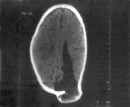
attenuation area of size 1.5 × l.2 cm of CSF density was seen protruding
through the gap on C.T. examination (Fig. 1). Widening of the
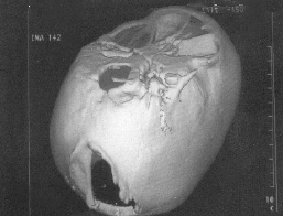
sulcal spaces in the left parietal region was also seen. A three
dimensional reconstruction was done to demonstrate the defect for the
operating surgeon (Fig. 2).
 |
 |
|
Fig. 1. Axial CT section showing defect
in the skull with a cystic density lesion pushing through and
communicating with the left lateral ventricle. Also noted is
widening of the sulcal spaces. |
Fig. 2. Three dimensional reconstruction
to demons-trate the calvarial defect.
|
The patient was operated upon and corrective surgery
was done. There were no intra-operative or post-operative complica-tions
and the patient was doing fine on the follow up visit a month after the
operation.
Discussion
Growing skull fractures usually occur after severe
head trauma during the first three years of the life (particularly
infancy) and almost never after 8 yrs of life. The incidence reported is
only 0.05 to 0.1% of skull fractures in childhood(1,2).
During this stage, the brain volume increases
rapidly, which is in part responsible for its development. Though the
development of growing skull fractures is multifactorial, the
predominant factor in their causation is the presence of lacerated
duramater. The pulsatile force, of the brain during its growth causes
the fracture in the thin skull to enlarge. This interposition of tissue
prevents osteoblasts from migrating to the fracture site, inhibiting
healing. The resorption of the adjacent bone by the continuous pressure
from tissue herniation through the bone gap adds to the progression of
the fracture line.
The brain extrusion may be present shortly after
diastatic linear fracture in neonates and young infants resulting in
focal dilation of the lateral ventricle near the growing fracture(3).
This dilatation is reversible and may normalize after surgical
repair(4). Cranial defects have been found never to increase in size if
the underlying dura is intact. Also, craniotomies performed without
watertight closure of dural lacerations have been found to lead to
growing skull fractures. These support dural tear as being the major
risk factor in the development of a growing skull fracture.
Another risk factor is severity of the underlying
trauma. A linear fracture associated with hemorrhagic contusion of
subjacent brain suggests a trauma significant enough to cause dural
laceration. The brain at the growing fracture site shows a
cerebromeningeal cicatrix formation. Cystic changes at the growing
fracture site may be because of cystic encephalomalacia. Post traumatic
aneurysms and subdural hematomas have also been reported to accompany
growing skull fractures(5,6). Though most patients show damage to
underlying brain, this finding is not a prerequisite for the development
of growing skull fractures(7).
These skull fractures, after reaching their maximum
extent, cease to grow and remain stable throughout adulthood(2). A
depressed fracture usually does not become a growing fracture(8) but a
linear fracture extending from a depressed one can become one(9). A
fracture with a diastasis of >4 mm may be considered at risk of
developing a growing skull fracture(3,10). But a post traumatic
diastasis of a cranial suture is an unusual site for a growing fracture.
The usual site is the parietal region. A growing fracture at the skull
base may present with ocular proptosis or CSF rhinorrhea or otorrhea.
Owing to the risk of neurological deterioration and
development of seizure disorders, surgical correction of growing
fractures is recommended.
Contributors: SGI was responsible for the image
reconstruction, literature search and preparation of the manuscript. PS
was responsible for the drafting of the manuscript. GDK critically
evaluated the paper and gave the final approval of the version of the
manuscript to be sent for publishing.
Funding: None.
Competing interests: None stated.
